Review
. 2018 Jan;28(1):10-21.
doi: 10.1016/j.tcm.2017.07.004. Epub 2017 Jul 13.

Cardiac amyloidosis: An update on pathophysiology, diagnosis, and treatment

Affiliations
Review

Cardiac amyloidosis: An update on pathophysiology, diagnosis, and treatment

Omar K Siddiqi et al. Trends Cardiovasc Med. 2018 Jan.

Abstract

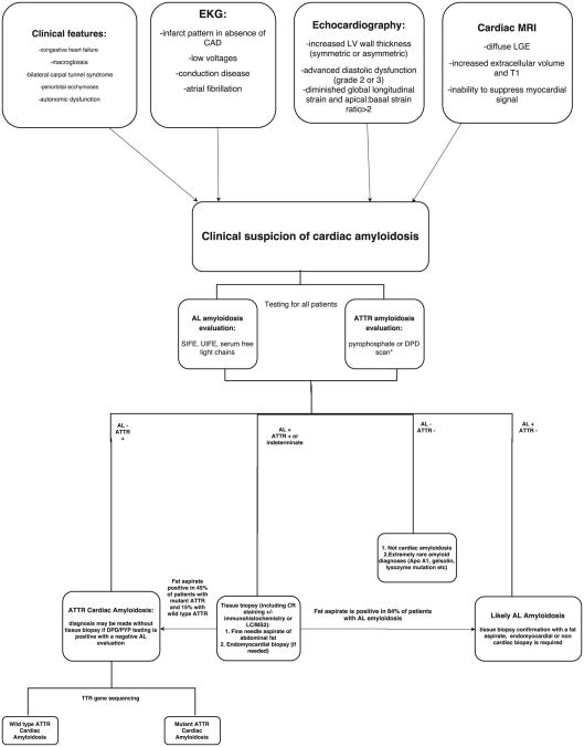
The amyloidoses are a group of systemic diseases characterized by organ deposition of misfolded protein fragments of diverse origins. The natural history of the disease, involvement of other organs, and treatment options vary significantly based on the protein of origin. In AL amyloidosis, amyloid protein is derived from immunoglobulin light chains, and most often involves the kidneys and the heart. ATTR amyloidosis is categorized as mutant or wild-type depending on the genetic sequence of the transthyretin (TTR) protein produced by the liver. Wild-type ATTR amyloidosis mainly involves the heart, although the reported occurrence of bilateral carpal tunnel syndrome, spinal stenosis and biceps tendon rupture in these patients speaks to more generalized protein deposition. Mutant TTR is marked by cardiac and/or peripheral nervous system involvement. Cardiac involvement is associated with symptoms of heart failure, and dictates the clinical course of the disease. Cardiac amyloidosis can be diagnosed noninvasively by echocardiography, cardiac MRI, or nuclear scintigraphy. Endomyocardial biopsy may be needed in the case of equivocal imaging findings or discordant data. Treatment is aimed at relieving congestive symptoms and targeting the underlying amyloidogenic process. This includes anti-plasma cell therapy in AL amyloidosis, and stabilization of the TTR tetramer or inhibition of TTR protein production in ATTR amyloidosis. Cardiac transplantation can be considered in highly selected patients in tandem with therapy aimed at suppressing the amyloidogenic process, and appears associated with durable long-term survival.

Keywords: AL amyloidosis; Restrictive cardiomyopathy; Transthyretin amyloidosis.


Figures

Figure 1
Figure 1
a-h. Echocardiography characteristics of cardiac amyloidosis. Parasternal long- and short-axis views demonstrating severely increased LV wall thickness, with granular sparkling, which may be seen in cardiac amyloidosis. Pericardial effusions are common (1a-b). Atrial dilation reflects elevated filling pressures, as well as atrial infiltration of amyloid protein (1c). Elevated RV wall thickness (> 5mm) is often seen as well (1d). Longitudinal strain in apical 4-, 2- and 3-chamber views showing severely reduced global longitudinal strain (GLS) of -12.5% with an apical to basal strain ratio of 2.7 (1e-h).
Figure 1
Figure 1
a-h. Echocardiography characteristics of cardiac amyloidosis. Parasternal long- and short-axis views demonstrating severely increased LV wall thickness, with granular sparkling, which may be seen in cardiac amyloidosis. Pericardial effusions are common (1a-b). Atrial dilation reflects elevated filling pressures, as well as atrial infiltration of amyloid protein (1c). Elevated RV wall thickness (> 5mm) is often seen as well (1d). Longitudinal strain in apical 4-, 2- and 3-chamber views showing severely reduced global longitudinal strain (GLS) of -12.5% with an apical to basal strain ratio of 2.7 (1e-h).
Figure 2
Figure 2
a-c. Cardiac MRI demonstrating characteristic late gadolinium enhancement (LGE) patterns in cardiac amyloidosis. Phase sensitive, inversion recovery (PSIR) LGE images are illustrated in the 4-chamber (2a) and short axis (2b and 2c) views, in different patients with light chain (AL) amyloidosis.
Figure 2
Figure 2
a-c. Cardiac MRI demonstrating characteristic late gadolinium enhancement (LGE) patterns in cardiac amyloidosis. Phase sensitive, inversion recovery (PSIR) LGE images are illustrated in the 4-chamber (2a) and short axis (2b and 2c) views, in different patients with light chain (AL) amyloidosis.
Figure 3
Figure 3
a-d. Tc99m pyrophosphate (PYP) scans in AP and lateral projections revealing equivocal (Grade 1) uptake (3a and 3b) in a patient with AL amyloidosis, and strongly positive (Grade 3) uptake (3c and 3d) by the heart when compared to the surrounding ribs in a patient with ATTR wild-type amyloidosis.
Figure 3
Figure 3
a-d. Tc99m pyrophosphate (PYP) scans in AP and lateral projections revealing equivocal (Grade 1) uptake (3a and 3b) in a patient with AL amyloidosis, and strongly positive (Grade 3) uptake (3c and 3d) by the heart when compared to the surrounding ribs in a patient with ATTR wild-type amyloidosis.
Figure 4
Figure 4
Suggested algorithm for the diagnosis of cardiac amyloidosis, and for differentiating between ATTRwt, ATTRm, and AL cardiac amyloidosis. Adapted from Siddiqi OK, Ruberg FL. Challenging the Myths of Cardiac Amyloidosis. European Heart Journal 2017 (64).

Comment in

References

    1. Sipe JD, Cohen AS. Review: history of the amyloid fibril. J Struct Biol. 2000;130(2-3):88–98. - PubMed
    1. Falk RH, Alexander KM, Liao R, Dorbala S. AL (Light-Chain) Cardiac Amyloidosis: A Review of Diagnosis and Therapy. Journal of the American College of Cardiology. 2016;68(12):1323–41. - PubMed
    1. Gertz MA, Comenzo R, Falk RH, Fermand JP, Hazenberg BP, Hawkins PN, et al. Definition of organ involvement and treatment response in immunoglobulin light chain amyloidosis (AL): a consensus opinion from the 10th International Symposium on Amyloid and Amyloidosis, Tours, France, 18-22 April 2004. American journal of hematology. 2005;79(4):319–28. - PubMed
    1. Skinner M, Cohen AS. The prealbumin nature of the amyloid protein in familial amyloid polyneuropathy (FAP)-swedish variety. Biochemical and biophysical research communications. 1981;99(4):1326–32. - PubMed
    1. Costa PP, Figueira AS, Bravo FR. Amyloid fibril protein related to prealbumin in familial amyloidotic polyneuropathy. Proceedings of the National Academy of Sciences of the United States of America. 1978;75(9):4499–503. - PMC - PubMed

Publication types

MeSH terms

Substances

Supplementary concepts