Case Reports
. 2017 Jul 24:2017:bcr2017220645.
doi: 10.1136/bcr-2017-220645.

Case of a strangulated right paraduodenal fossa hernia in a malrotated gut

Affiliations
Case Reports

Case of a strangulated right paraduodenal fossa hernia in a malrotated gut

Michelle Ong et al. BMJ Case Rep. .

Abstract

We report an unusual case of a strangulated internal hernia resulting from a right paraduodenal fossa hernia (PDH) in the context of bowel malrotation. There are few documented cases of PDHs associated with a concomitant gut malrotation. Emergency laparotomy was performed based on clinical and radiological. Intraoperatively, the proximal jejunum was seen to enter a hernia sac formed by an aberrant duodenojejunal flexure located to the right of the aorta. This was presumed to be a strangulated internal hernia of the paraduodenal recess in a malrotated gut. The hernia neck was widened and the sac obliterated to allow reduction of the contents. On reduction and warming, the insulted small bowel appeared viable and returned to the abdominal cavity without resection.

Keywords: gastroenterology; gastrointestinal system; stomach and duodenum.


Conflict of interest statement

Competing interests: None declared.

Figures
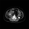
Figure 1
Figure 1
Axial view of CT imaging with intravenous contrast acquired in the arterial phase demonstrating SB contained within an internal sac on the right side of the abdomen. SB within sac is poorly enhancing with localised oedema. SB, small bowel.
Figure 2
Figure 2
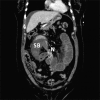
Coronal view CT imaging with intravenous contrast acquired in the portal venous phase demonstrating the neck of the hernia sac (N) and resulting closed-loop obstruction of the small intestine (SB). Other identifiable features include dilated proximal SB, perihepatic and lower abdominal free fluid and nasogastric tube located in the stomach. N, neck; SB, small bowel.
Figure 3
Figure 3
Sagittal view of CT imaging with intravenous contrast acquired in the arterial phase demonstrating the absence of the duodenum crossing the abdominal aorta (A) inferior to the root of the superior mesenteric artery. The absence of the duodenum to in the left of the abdomen suggests a bowel malrotation, resulting in the duodenojejunal flexure located on the right side. Dilated proximal and collapsed distal SB, as well as free pelvic fluid, are also seen. SB, small bowel.

References

    1. Kuzinkovas V, Haghighi K, Singhal R, et al. . Paraduodenal hernia: a rare cause of abdominal pain. Can J Surg 2008;51:E127–E128. - PMC - PubMed
    1. Virich G, Davies W. A massive left paraduodenal Fossa hernia as an unusual cause of small bowel obstruction. Ann R Coll Surg Engl 2010;92:e7–e9. 10.1308/147870810X12659688851997 - DOI - PMC - PubMed
    1. Singh S, Das A, Chawla AS, et al. . A rare presentation of midgut malrotation as an acute intestinal obstruction in an adult: two case reports and literature review. Int J Surg Case Rep 2013;4:72–5. 10.1016/j.ijscr.2012.10.005 - DOI - PMC - PubMed
    1. Rajesh S, Kumar PS, Maheshwari G, et al. . Malrotation of small bowel-diagnostic computed tomography (CT) signs and intraoperative findings. Indian J Surg 2015;77:600–2. 10.1007/s12262-013-0938-9 - DOI - PMC - PubMed
    1. Bittner JG, Edwards MA, Harrison SJ, et al. . Laparoscopic repair of a right paraduodenal hernia. JSLS 2009;13:242–9. - PMC - PubMed

Publication types

Supplementary concepts