. 2017 Aug 8;114(32):E6536-E6545.
doi: 10.1073/pnas.1704782114. Epub 2017 Jul 24.

Unique structural features of the AIPL1-FKBP domain that support prenyl lipid binding and underlie protein malfunction in blindness

Affiliations

Unique structural features of the AIPL1-FKBP domain that support prenyl lipid binding and underlie protein malfunction in blindness

Ravi P Yadav et al. Proc Natl Acad Sci U S A. .

Abstract

FKBP-domain proteins (FKBPs) are pivotal modulators of cellular signaling, protein folding, and gene transcription. Aryl hydrocarbon receptor-interacting protein-like 1 (AIPL1) is a distinctive member of the FKBP superfamily in terms of its biochemical properties, and it plays an important biological role as a chaperone of phosphodiesterase 6 (PDE6), an effector enzyme of the visual transduction cascade. Malfunction of mutant AIPL1 proteins triggers a severe form of Leber congenital amaurosis and leads to blindness. The mechanism underlying the chaperone activity of AIPL1 is largely unknown, but involves the binding of isoprenyl groups on PDE6 to the FKBP domain of AIPL1. We solved the crystal structures of the AIPL1-FKBP domain and its pathogenic mutant V71F, both in the apo form and in complex with isoprenyl moieties. These structures reveal a module for lipid binding that is unparalleled within the FKBP superfamily. The prenyl binding is enabled by a unique "loop-out" conformation of the β4-α1 loop and a conformational "flip-out" switch of the key W72 residue. A second major conformation of apo AIPL1-FKBP was identified by NMR studies. This conformation, wherein W72 flips into the ligand-binding pocket and renders the protein incapable of prenyl binding, is supported by molecular dynamics simulations and appears to underlie the pathogenicity of the V71F mutant. Our findings offer critical insights into the mechanisms that underlie AIPL1 function in health and disease, and highlight the structural and functional diversity of the FKBPs.

Keywords: AIPL1; FKBP; PDE6; chaperone; photoreceptor.


Conflict of interest statement

The authors declare no conflict of interest.

Figures

Fig. 1.
Fig. 1.
Comparison of FKBP-domain structures of AIPL1, AIP, and FKBP12. (A) AIPL1 (PDB ID code 5U9A). Highlighted are the 57-residue insert region (blue), which forms three α helices α2, α3, and α4); the β4-α1 loop (cyan), which is in the loop-out conformation; and the N-terminal α0 region (orange), which is disordered. (B) AIP (PDB ID code 2LKN). Highlighted are the 57-residue insert region (faded green), whose α3 helix region is disordered; the β4-α1 loop (bright green), which is in a loop-in conformation; and the α0 region (yellow), which is structured. (C) FKBP12 (PDB ID code 1FKB). Highlighted are the 20-residue hairpin loop counterpart to the insert region in AIPL1 and AIP (salmon); the β4-α1 loop (magenta), which is in a loop semi-in conformation; and rapamycin (indicated in purple stick representation). (D) Sequence alignment of human FKBP12 with the FKBP-domains of AIP and AIPL1. The key elements of the protein secondary structure are identified. A single-turn α-helix between the β3 and β4 strands is denoted as αt. Arrowheads indicate AIPL1 residues V71 (mutated in LCA) and W72 (flips between two conformations, as detected by NMR). Arrows indicate residues in the β4-α1 loop that differ between AIPL1 and AIP, and contribute to the loop-out or loop-in conformation. Dashed underline highlights the α0-helix region, which is α-helical in AIP but not in AIPL1. The well-ordered α3 helix in AIPL1 corresponds to the poorly ordered flexible region in AIP.
Fig. 2.
Fig. 2.
Hydrophobic cavity access and the role of the β4-α1 loop and the α1 tryptophan residue. (A and B) The FKBP domains of AIP and AIPL1 shown in surface representation. In AIP, the hydrophobic cavity is sealed from the surface because of the loop-in conformation of β4-α1 (A), but in AIPL1 the loop-out conformation of β4-α1 provides two entrances to the cavity (B). (C) Comparison of the β4-α1 loop, showing that irrespective of the presence or absence of ligands, in most FKBP proteins it is in the loop semi-in conformation (magenta; PDB ID codes 1C9H, 1FKB, 1Q1C, 2PBC, 2PPN, 2VN1, 3JYM), in AIP it is in the loop-in conformation (green; PDB ID code 2LKN), and in AIPL1 it is in the loop-out conformation (cyan; PDB ID codes 5U9A, 5U9I, 5U9J). In AIP, the side chain of W73 (green sticks) is flipped into the cavity and forms its base, whereas in AIPL1 W72 (cyan sticks) is flipped out and thus the cavity is deeper.
Fig. 3.
Fig. 3.
FC- and GGpp-bound structures of AIPL1. (A and B) The farnesyl moiety of FC (yellow) (A), and the geranylgeranyl moiety of GGpp (pink) (B) penetrate the hydrophobic binding pocket, with the latter sitting deeper. In both cases, W72 (cyan) is flipped out, and thus the ligands can enter the cavity. (C) Binding of FC-FITC to the AIP–FKBP domain, the AIPL1–FKBP domain, and two mutant forms of the latter (N65K/M66K/E70P and V71F). Fluorescence polarization (mP) of FC-FITC is plotted as a function of concentration of the binding protein, and data are fit using a single-site binding equation. The results of a representative experiment are shown. The Kd values, calculated based on three experiments, are: 50 ± 6 nM for AIPL1–FKBP; 795 ± 65 nM for AIPL1–FKBP N65K/M66K/E70P; 101 ± 13 nM for AIPL1–FKBP V71F, t test vs. AIPL1–FKBP significance value P = 0.02; and >5,000 nM for AIP–FKBP.
Fig. 4.
Fig. 4.
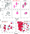
Apo AIPL1–FKBP exists in two conformations in solution that collapse to one conformation on FC binding. (A) The M79, A85, and Ι151 amide regions of the 15N/1H HSQC spectra of AIPL1–FKBP in the absence and presence of FC and their spectra overlays. (B and C) Overlay of 13C/1H HSQC spectra of AIPL1–FKBP in the absence and presence of FC. (B) Ile CδH3 region. (C) Val CγH3 and Leu CδH3 region. A subset of assigned peaks is labeled.
Fig. 5.
Fig. 5.
Titration of geraniol into AIPL1–FKBP WT and L100Ι mutant. Shown are overlays of the Val CγH3 and Leu CδH3 region of 13C/1H HSQC spectra of AIPL1–FKBP WT and L100Ι at the indicated geraniol concentrations. (A and B) AIPL1–FKBP WT. (C) AIPL1–FKBP L100Ι. Protein concentration used in these experiments is 140 µM. (D) Ribbon plot of the apo AIPL1–FKBP crystal structure. W72 and L100 are shown in sticks. W72 in magenta indicates the flip-out conformation that was detected in the crystal structure, and W72 in green indicates the flip-in conformation as deduced from the NMR data. The ribbon in cyan indicates the region encompassing residues G64-L76 (the β4-α1 loop plus part of α1 helix), whose backbone amides are broad beyond detection in the AIPL1–FKBP Δ111–132:FC complex.
Fig. 6.
Fig. 6.
AIPL1–FKBP V71F has a flip-in major conformation in solution. (A and B) Overlays of 13C/1H HSQC spectra of the Ile CδH3 region of AIPL1–FKBP WT and the V71F mutant in the absence and presence of FC, respectively. (C) Overlap of the spectra of AIPL1–FKBP V71F in the absence and presence of FC. (D) Ribbon plot of the AIPL1–FKBP V71F apo crystal structure, zoomed around V71F. Selected residues are shown in magenta sticks. The NMR data suggest that in the apo protein in solution, the W72 indole ring has rotated into the FC-binding pocket (indicated by yellow stick). The ribbon in cyan indicates the region encompassing residues G64-L76 (the β4-α1 loop plus part of α1 helix), whose backbone amides are broad beyond detection in the AIPL1–FKBPΔ111–132:FC complex.
Fig. 7.
Fig. 7.
MD simulations of apo AIPL1–FKBP and V71F. (A) Typical WT AIPL1–FKBP run (run 2 in SI Appendix, Table S1) (initial model in slate; model at end of 183.75-ns MD simulation in cyan). (B) Typical V71F run (run 3; initial model in white; model at end of 206.75 ns in wheat). (C) Atypical V71F run (run 2; initial model in green; model at end of 120.25-ns MD simulation in magenta). W72 remains in the flipped-out position in all MD runs of WT AIPL1–FKBP (A) and V71F (B), except in the MD run V71F-2 (C), where movement of F71 caused a clash with W72 and resulted in its immediate flip-in where it remained until the end of the simulation (120 ns). (D) The equivalent W73 in AIP was found to be in the flipped-in position. The β4-α1 loop remains in the loop-out position for all WT AIPL1–FKBP and V71F MD runs, suggesting that the flip-in position of W72 may not be strongly coupled to the loop-in β4-α1 loop conformation. (E) Root mean square fluctuations for residues in 5 WT FKBP–AIPL1 and 7 V71F independent MD simulations indicating that α3 is the most flexible region. (F) Movement of α3 during MD simulations for WT FKBP–AIPL1 from the starting position (cyan) toward the more commonly seen lid-closed (red) position than the lid-open (blue) position seen only in WT-1 simulation (SI Appendix, Table S1). AIP’s equivalent α3 region is in yellow. (G) FKBP–AIPL1 WT structure at the beginning (cyan) and (H) after about 180 ns (red) of a MD simulation (WT-2) (SI Appendix, Table S1). As the simulation progresses, the entrance to the hydrophobic cavity is blocked by W72 (yellow), which lies near the beginning of α1 and W116 (magenta) which lies near the beginning of α3.

References

    1. Tong M, Jiang Y. FK506-binding proteins and their diverse functions. Curr Mol Pharmacol. 2015;9:48–65. - PMC - PubMed
    1. Hausch F. FKBPs and their role in neuronal signaling. Biochim Biophys Acta. 2015;1850:2035–2040. - PubMed
    1. Storer CL, Dickey CA, Galigniana MD, Rein T, Cox MB. FKBP51 and FKBP52 in signaling and disease. Trends Endocrinol Metab. 2011;22:481–490. - PMC - PubMed
    1. MacMillan D. FK506 binding proteins: Cellular regulators of intracellular Ca2+ signalling. Eur J Pharmacol. 2013;700:181–193. - PubMed
    1. Van Duyne GD, Standaert RF, Karplus PA, Schreiber SL, Clardy J. Atomic structures of the human immunophilin FKBP-12 complexes with FK506 and rapamycin. J Mol Biol. 1993;229:105–124. - PubMed

Publication types

MeSH terms

LinkOut - more resources