. 2017 Aug 8;114(32):E6623-E6631.
doi: 10.1073/pnas.1706055114. Epub 2017 Jul 24.

Therapeutically targeting glypican-2 via single-domain antibody-based chimeric antigen receptors and immunotoxins in neuroblastoma

Affiliations

Therapeutically targeting glypican-2 via single-domain antibody-based chimeric antigen receptors and immunotoxins in neuroblastoma

Nan Li et al. Proc Natl Acad Sci U S A. .

Abstract

Neuroblastoma is a childhood cancer that is fatal in almost half of patients despite intense multimodality treatment. This cancer is derived from neuroendocrine tissue located in the sympathetic nervous system. Glypican-2 (GPC2) is a cell surface heparan sulfate proteoglycan that is important for neuronal cell adhesion and neurite outgrowth. In this study, we find that GPC2 protein is highly expressed in about half of neuroblastoma cases and that high GPC2 expression correlates with poor overall survival compared with patients with low GPC2 expression. We demonstrate that silencing of GPC2 by CRISPR-Cas9 or siRNA results in the inhibition of neuroblastoma tumor cell growth. GPC2 silencing inactivates Wnt/β-catenin signaling and reduces the expression of the target gene N-Myc, an oncogenic driver of neuroblastoma tumorigenesis. We have isolated human single-domain antibodies specific for GPC2 by phage display technology and found that the single-domain antibodies can inhibit active β-catenin signaling by disrupting the interaction of GPC2 and Wnt3a. To explore GPC2 as a potential target in neuroblastoma, we have developed two forms of antibody therapeutics, immunotoxins and chimeric antigen receptor (CAR) T cells. Immunotoxin treatment was demonstrated to inhibit neuroblastoma growth in mice. CAR T cells targeting GPC2 eliminated tumors in a disseminated neuroblastoma mouse model where tumor metastasis had spread to multiple clinically relevant sites, including spine, skull, legs, and pelvis. This study suggests GPC2 as a promising therapeutic target in neuroblastoma.

Keywords: chimeric antigen receptor T-cell therapy; glypican; immunotoxin; neuroblastoma; single-domain antibody.


Conflict of interest statement

The authors declare no conflict of interest.

Figures
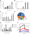
Fig. 1.
Fig. 1.
Isolation of GPC2-specific human single-domain antibodies by phage display. (A) Phage-displayed single-domain antibody fragments were selected against recombinant GPC2–hFc after four rounds of panning. A gradual increase in phage titers was observed during each round of panning. (B) Polyclonal phage ELISA from the output phage of each round of panning. BSA was used as an irrelevant antigen. (C) Monoclonal phage ELISA of the seven GPC2 binders. (D) Distribution of unique sequences of GPC2 binders in 27 selected phage clones. (E) Monoclonal phage ELISA analysis of cross-reactivity of GPC2 binders to human GPC1 and GPC3 and mouse GPC2. (F) Octet association and dissociation kinetic analysis for the interaction between various concentrations of the LH7 antibody and human GPC2. All data are represented as mean ± SEM of three independent experiments.
Fig. 2.
Fig. 2.
GPC2 expression in human neuroblastoma tumors and normal human tissues. (A) GPC2 protein levels in human neuroblastoma cell lines, including SKNSH, LAN1, IMR5, LAN5, IMR32, and NBEB as determined by Western blotting. (B) Expression of GPC2 in neuroblastoma tumors (iiv) and normal nerve (v and vi) tissues as determined by immunohistochemistry. (C) Expression of GPC2 in human normal tissues, including nerve, brain, heart, lung, liver, stomach, small intestine, colon, pancreas, spleen, kidney, and thyroid as determined by immunohistochemistry. The tissues were labeled with 1 μg/mL LH7–mFc antibody. The final magnification of all images was 100×. (D) Kaplan–Meier analysis of overall survival in patients with neuroblastoma with high GPC2 mRNA expression (n = 18) and low GPC2 mRNA expression (n = 458) from the Kocak dataset in the R2 Genomics Analysis and Visualization Platform. (E) Kaplan–Meier analysis of event-free survival in patients with neuroblastoma with high GPC2 mRNA expression (n = 20) and low GPC2 mRNA expression (n = 456) from the Kocak dataset.
Fig. 3.
Fig. 3.
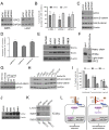
Genetic silencing of GPC2 inhibits neuroblastoma tumor cell growth and induces apoptosis by suppressing Wnt/β-catenin signaling. (A) GPC2 protein expression in LAN1 and IMR5 neuroblastoma cells after siRNA-mediated knockdown of GPC2. (B) Inhibition of tumor cell growth by GPC2 siRNAs in both LAN1 and IMR5 cell lines. (C) GPC2 expression in IMR5 neuroblastoma cells after GPC2 KO using the CRISPR-Cas9 technique. GPC2 KO decreased active β-catenin protein levels at 72 h posttransfection. (D) Caspase 3/7 activity in IMR5 cells after treatment with GPC2 targeted sgRNA. (E) Protein expression of Wnt3a and Wnt11 in neuroblastoma cell lines. (F) Interaction between GPC2 and Wnt3a as determined by immunoprecipitation. (G) Reduction of active β-catenin levels by LH7 treatment after 6 h in HEK-293 SuperTopFlash cells that were stimulated with Wnt3a CM. (H) LH7 suppressed the expression of β-catenin in HEK-293 SuperTopFlash cells that were stimulated with LiCl and/or Wnt3a CM. Whole cell lysates were collected after 6 h of treatment. (I) The anti-GPC2 antibodies decreased TopFlash activity in Wnt3a-activated HEK-293 SuperTopFlash cells after 6 h of treatment. (J) N-Myc protein level in neuroblastoma cell lines as determined by Western blotting. (K) Inhibition of N-Myc expression by silencing GPC2 in neuroblastoma cells. (L) The proposed mechanism mediated by anti-GPC2 antibodies to inhibit neuroblastoma cell growth. Blockade of GPC2 suppresses the expression of β-catenin and its targeted genes, including N-Myc. All data are represented as mean ± SEM of three independent experiments. *P < 0.05, **P < 0.01.
Fig. 4.
Fig. 4.
Recombinant immunotoxins against GPC2 inhibit neuroblastoma tumor growth in vitro and in vivo. (A) Purity of LH1–PE38 (molecular weight of 53 kDa), LH4–PE38 (molecular weight of 52 kDa), and LH7–PE38 (molecular weight of 52 kDa) as determined by SDS/PAGE. (BD) Effectiveness of anti-GPC2 immunotoxins on the growth of IMR5 (B), LAN1 (C), and SKNSH (D) cell lines, as measured by the WST-8 assay. An antimesothelin immunotoxin was used as an irrelevant control immunotoxin. (E) Toxicity detection of LH7–PE38 in vivo. Athymic nu/nu nude mice were treated with indicated doses of immunotoxin i.v. every other day for a total of 10 injections. Each arrow indicates an individual injection (n = 5 per group). (F) Antitumor activity of LH7–PE38. Athymic nu/nu nude mice were s.c. inoculated with 1 × 10⁷ LAN1 cells mixed with Matrigel. When tumors reached an average volume of 150 mm3, mice were treated with a 0.4-mg/kg dose of LH7–PE38 i.v. every other day for 10 injections. Each arrow indicates an individual injection; n = 5 per group. *P < 0.05. (G) Body weight of the mice treated in F. (H) Representative pictures of tumors from control and treated mice at the end of study. Values represent mean ± SEM. IT, immunotoxin.
Fig. 5.
Fig. 5.
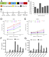
The CAR T cells targeting GPC2 kill neuroblastoma cells. (A) Schematic diagram of bicistronic lentiviral constructs expressing CARs targeting GPC2 along with GFP using the T2A ribosomal skipping sequence. (B) Timeline of CAR T-cell production. (C) GPC2-specific CAR expression on human T cells transduced with lentiviral particles was analyzed using flow cytometry by detection of GFP fluorescence. (DE) Cytolytic activities of GPC2 targeting CAR T cells in cell assays. The luciferase-expressing IMR5 (D) and SKNSH (E) neuroblastoma cells were cocultured with mock or GPC2 CAR-transduced T cells at the indicated effector (E):target (T) ratios for 20 h, and specific lysis was measured using a luminescent-based cytolytic assay. (FG) The above culture supernatants at an E:T ratio of eight were harvested to measure IFN-γ (F) and TNF-α (G) secretions via ELISA. All data are represented as mean ± SEM of three independent experiments. *P < 0.05, **P < 0.01.
Fig. 6.
Fig. 6.
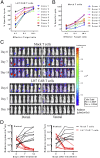
GPC2-specific CAR T cells demonstrate potent activity in mice bearing human neuroblastomas. (AB) Cytotoxic activity of LH7 CAR T cells derived from multiple donors. PMBCs were isolated from eight healthy donors. The luciferase-expressing IMR5 cells were cocultured with LH7 CAR-transduced T cells (A) or mock T cells (B) at the indicated effector (E):target (T) ratios for 20 h, and specific lysis was measured using a luminescent-based cytolytic assay. (C) LH7 CART demonstrated potent antitumor activity by suppressing metastatic IMR5 neuroblastoma cells as measured by bioluminescent imaging. Animals (n = 8 per group) were i.v. injected with a single infusion of 30 × 106 mock T cells or LH7 CAR T cells. (D) Quantitation of bioluminescence in mice treated in C. Values represent mean ± SEM.

References

    1. Maris JM, Hogarty MD, Bagatell R, Cohn SL. Neuroblastoma. Lancet. 2007;369:2106–2120. - PubMed
    1. Brodeur GM. Neuroblastoma: Biological insights into a clinical enigma. Nat Rev Cancer. 2003;3:203–216. - PubMed
    1. Yu AL, et al. Children’s Oncology Group Anti-GD2 antibody with GM-CSF, interleukin-2, and isotretinoin for neuroblastoma. N Engl J Med. 2010;363:1324–1334. - PMC - PubMed
    1. Matthay KK, et al. Children’s Cancer Group Treatment of high-risk neuroblastoma with intensive chemotherapy, radiotherapy, autologous bone marrow transplantation, and 13-cis-retinoic acid. N Engl J Med. 1999;341:1165–1173. - PubMed
    1. Orentas RJ, et al. Identification of cell surface proteins as potential immunotherapy targets in 12 pediatric cancers. Front Oncol. 2012;2:194. - PMC - PubMed

Publication types

MeSH terms