. 2017 Jul 24;7(1):6228.
doi: 10.1038/s41598-017-05875-z.

CDKL5 localizes at the centrosome and midbody and is required for faithful cell division

Affiliations

CDKL5 localizes at the centrosome and midbody and is required for faithful cell division

Isabella Barbiero et al. Sci Rep. .

Abstract

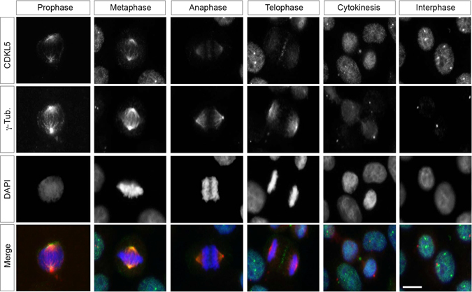
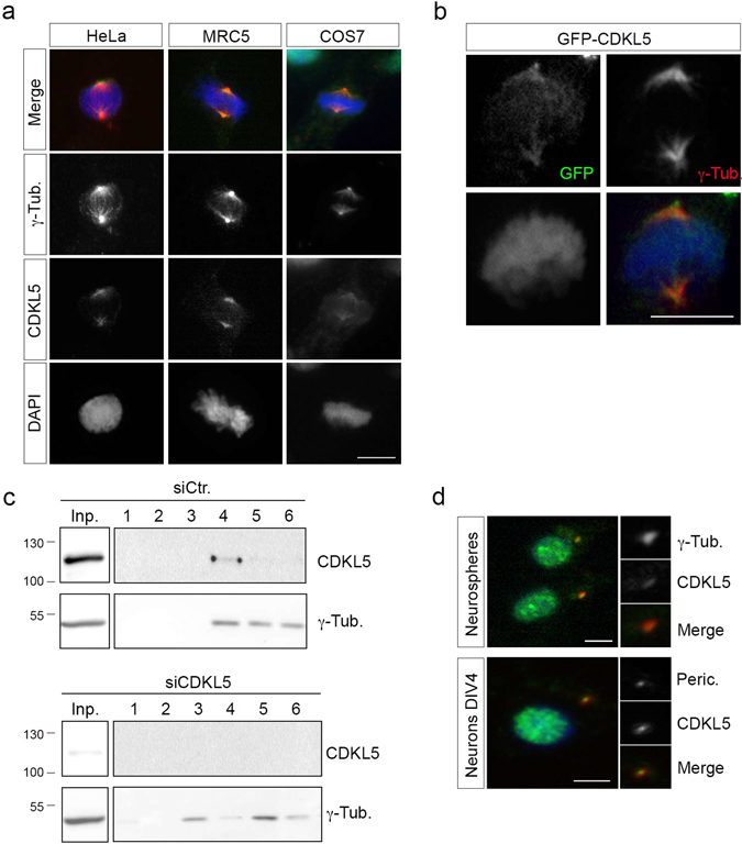
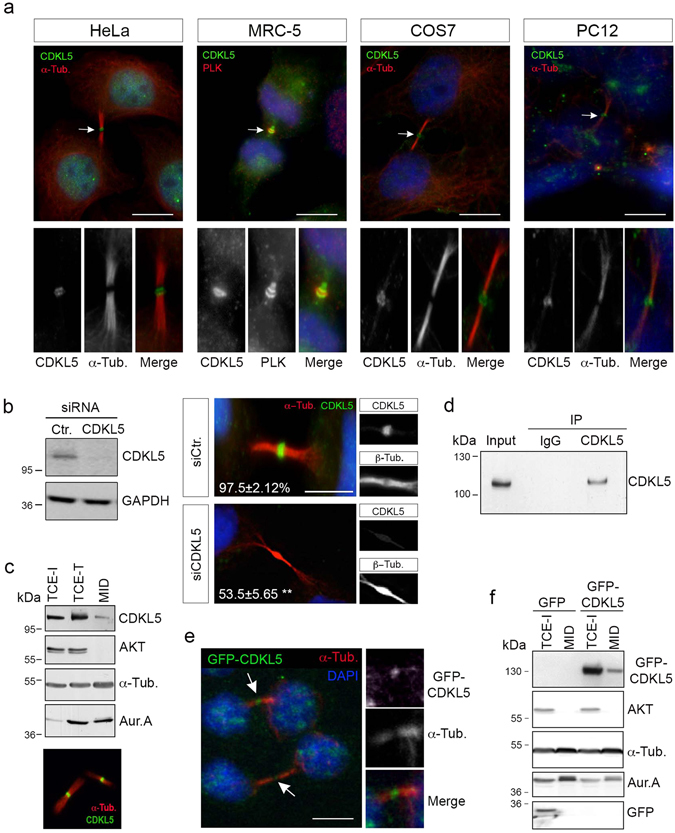
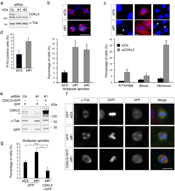
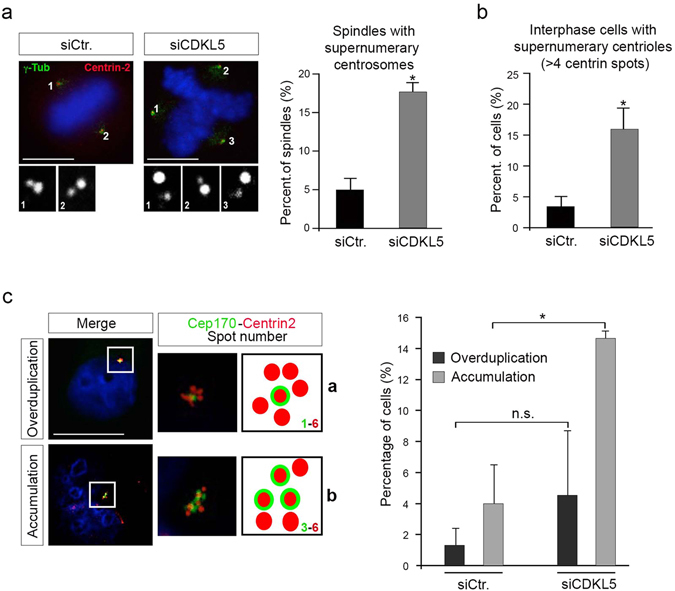
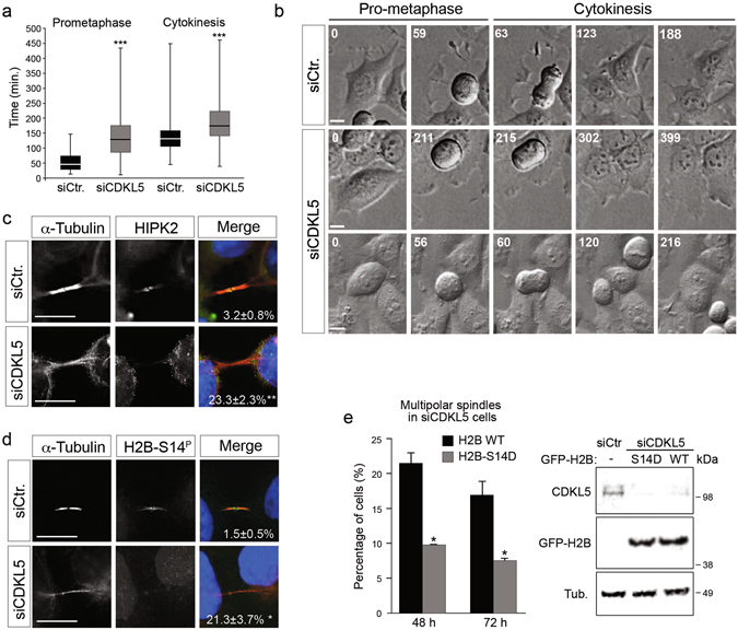
The cyclin-dependent kinase-like 5 (CDKL5) gene has been associated with rare neurodevelopmental disorders characterized by the early onset of seizures and intellectual disability. The CDKL5 protein is widely expressed in most tissues and cells with both nuclear and cytoplasmic localization. In post-mitotic neurons CDKL5 is mainly involved in dendritic arborization, axon outgrowth, and spine formation while in proliferating cells its function is still largely unknown. Here, we report that CDKL5 localizes at the centrosome and at the midbody in proliferating cells. Acute inactivation of CDKL5 by RNA interference (RNAi) leads to multipolar spindle formation, cytokinesis failure and centrosome accumulation. At the molecular level, we observed that, among the several midbody components we analyzed, midbodies of CDKL5-depleted cells were devoid of HIPK2 and its cytokinesis target, the extrachromosomal histone H2B phosphorylated at S14. Of relevance, expression of the phosphomimetic mutant H2B-S14D, which is capable of overcoming cytokinesis failure in HIPK2-defective cells, was sufficient to rescue spindle multipolarity in CDKL5-depleted cells. Taken together, these results highlight a hitherto unknown role of CDKL5 in regulating faithful cell division by guaranteeing proper HIPK2/H2B functions at the midbody.


Conflict of interest statement

The authors declare that they have no competing interests.

Figures

Figure 1
Figure 1
CDKL5 localizes at the centrosome and at the midbody in proliferating cells. HeLa cells were stained with Abs against CDKL5 (polyclonal, green) and γ-tubulin (red) and, to visualize DNA, with DAPI (blue). Scale bar, 10 µm.
Figure 2
Figure 2
CDKL5 is localized at the centrosome. (a) The indicated cells were stained as in Fig. 1. (b) Exogenously expressed GFP-CDKL5 (green) localizes at the γ-tubulin positive (red) centrosome in HeLa cells. DAPI staining (blue) was used to visualize DNA. (c) HeLa cells were transfected with siCDKL5#1 or a control siRNA (siCtr.); centrosomes were purified 60 h post-transfection and the obtained fractions analyzed by WB with Abs against CDKL5 and γ-tubulin. Input corresponds to approximately 0.6% of the whole cell extract before fractionation. Fractions 1 and 6 are the bottom and top ones, respectively (n = 3 independent experiments). Input and fractions 1–6 are shown as different exposures of the same membrane; full-length blots are presented in Supplementary Figure S7a. (d) Neurospheres (upper) and primary hippocampal neurons (DIV4; lower) were stained with Abs against CDKL5 (polyclonal, green) and either γ-tubulin or pericentrin (both red) and DAPI (blue). The panels to the right show the magnified centrosome. Scale bar, 10 µm.
Figure 3
Figure 3
CDKL5 localizes at the midbody. (a) The indicated cells were immunostained with Abs against CDKL5 (polyclonal, green) and α-tubulin or PLK-1 (red) to mark the midbody. DAPI staining (blue) was used to visualize DNA. Arrows indicate the CDKL5 positive midbodies, shown enlarged in the lower panels. (b) HeLa cells were treated as in Fig. 2c and silencing of CDKL5 expression was verified by western blotting 60 h post siRNA transfection (left). Immunofluorescence analysis (right) showed that only 53.5% ± 5.65 of siCDKL5-treated cells were positive for staining with CDKL5 Ab (polyclonal) at the midbody as opposed to 97.5% ± 2.12 of siCtr cells (n = 2; **p < 0.01; unpaired t-test; 160 midbodies analyzed per condition). Scale bar, 5 µm. (c) Total cell extracts of interphase (TCE-I) and telophase (TCE-T) cells were analyzed by WB together with extracts of purified midbodies (MID) with the indicated Abs. Midbody isolation was confirmed by immunofluorescence with Abs against CDKL5 and α-tubulin before extraction (lower panel), (n = 3). (d) WB showing immunoprecipitation of a midbody protein extract with polyclonal anti-CDKL5 or anti-IgG. Input corresponds to 10% of the midbody extract before the immunoprecipitation. (n = 2). (e) Exogenously expressed GFP-CDKL5 (green) could be detected at the midbody in transfected Hela cells. Arrows indicate GFP-CDKL5 at the midbody. Right panels show the magnified midbody. (f) Midbodies (MID) were purified from HeLa cells expressing GFP or GFP-CDKL5 and the extracted proteins were analyzed by WB with the indicated Abs together with an interphase total cell extract (TCE-I) (n = 2). Scale bars in a and e, 10 µm.
Figure 4
Figure 4
Silencing of CDKL5 is associated with multipolar spindle formation and chromosome segregation defects. (a) CDKL5 expression in HeLa cells 60 h after transfection with two different siRNAs targeting CDKL5 or a control siRNA. α-tubulin was used as loading control. The asterisk indicates an unspecific band (b,c) HeLa cells treated as in a were stained against α-tubulin and DAPI 60 h post-transfection. In b, the percentage of mitotic multipolar spindles is reported as mean ± S.E.M. (n = 3; *p < 0.05, ANOVA followed by Dunnet’s post hoc analysis). In c, the frequency of the indicated phenotypes is shown as mean ± S.E.M. (n = 2; *p < 0.05; **p < 0.01; unpaired t-test; approximately 1000 counted cells per condition). In b and c, representative images of the indicated cells are shown above the graphs with DAPI in blue and α-tubulin in red; scale bar, 10 µm. In c, arrows indicate a chromosome bridge and micronuclei. A/T = ana-/telo-phase. (d) HeLa cells were treated as in a and analyzed for phosphorylated histone H3 (P-H3) 60 h after silencing. The percentage of P-H3 positive cells was calculated in three independent experiments counting approximately 2000 cells (mean ± S.E.M., unpaired t-test). (e) CDKL5 was expressed in HeLa cells 60 h post-silencing by transfection of a bicistronic vector expressing also GFP. Vertical cropping was performed to show different exposures of the same membrane; full-length blots are shown in Supplementary Figure S7b. (f) Representative images of spindles in GFP-positive cells (green) treated as in e by staining against α-tubulin (red) and with DAPI (blue). Scale bar, 10 µm. (g) Graph showing number of cells with multipolar spindles upon CDKL5 re-expression in silenced cells. ≥ 60 counted cells per condition (n = 3; mean ± S.E.M, *p < 0.05; ANOVA followed by Dunnet’s post hoc analysis). n.s. = not statistically significant.
Figure 5
Figure 5
Spindle multipolarity in CDKL5 depleted cells is due to centrosome accumulation. (ac) HeLa cells were silenced as in Fig. 4a and analyzed by IF 60 h after transfection. In (a) cells were stained with Abs anti-γ-tubulin and anti-centrin-2 to mark the spindle poles and the centrioles, respectively. DAPI staining (blue) was used to visualize DNA. The insets below show the magnified centrin-2 signal in the correspondingly numbered poles. The percentage of spindles with supernumerary centrosomes is reported to the right as mean ± S.E.M. (n = 2; *p < 0.05; unpaired t-test; approximately 70 spindles were analyzed per condition). In (b) cells were immunostained with centrin-2 and the percentage of interphase cells with more than 4 centrioles was reported as mean ± S.E.M. (n = 3; *p < 0.05; unpaired t-test; approximately 100 cells were analyzed per condition). In c cells were stained with anti-cep170 (green) and anti-centrin-2 (red) Abs. DAPI staining (blue) was used to visualize DNA. Representative images of different siCDKL5 phenotypes are shown on the left. The percentage of cells with the indicated phenotypes is reported to the right as mean ± S.E.M (n = 2; *p < 0.05; unpaired t-test; approximately 200 cells were analyzed per condition). n.s. = not significant. Scale bar, 10 µm.
Figure 6
Figure 6
CDKL5 depletion is associated with cytokinesis failure and impaired HIPK2/H2B activity. (a) Indicated HeLa cells were analyzed 60 h post-silencing by time-lapse videomicroscopy. The duration of prometaphase and cytokinesis is reported in box plot graphs (***p < 0.001, unpaired t test). (b) Still images related to Supplementary Movies S1, S2 and S3, respectively upper, middle and lower panels. Representative still images of siCDKL5 cells with very long cytokinesis time (middle panels) and of siCDKL5 cells that fail cytokinesis and form binucleated cell (lower panels) are showed. Time is indicated in minutes. Scale bar, 10 µm. (c, d) Midbody localization of HIPK2 (c) and H2B phosphorylated at S14 (d; H2B-S14P) was analyzed in the indicated HeLa cells 60 h post-silencing by staining with Abs against α–tubulin (red) and HIPK2/H2B-S14P (green) and with DAPI (blue). The percentage of cells with HIPK2/ H2B-S14P negative midbodies is indicated for each condition (n = 2; mean ± S.E.M., *p < 0.05; **p < 0.01 unpaired t-test; approximately 80 midbodies were analyzed per condition). Scale bar, 10 µm. (e) Graph showing percentage of multipolar spindles in siCDKL5 cells expressing GFP-H2B WT or GFP-H2B-S14D 48 and 72 h after siRNA transfection (n = 2; *p < 0.05; unpaired t-test; approximately 80 cells were analyzed per condition). WB showing CDKL5 and GFP-H2B protein levels 72 h after siRNA transfection is reported.

Similar articles

Cited by

References

    1. Montini E, et al. Identification and characterization of a novel serine-threonine kinase gene from the Xp22 region. Genomics. 1998;51:427–433. doi: 10.1006/geno.1998.5391. - DOI - PubMed
    1. Kilstrup-Nielsen C, et al. What we know and would like to know about CDKL5 and its involvement in epileptic encephalopathy. Neural Plast. 2012;2012:728267. doi: 10.1155/2012/728267. - DOI - PMC - PubMed
    1. Wang IT, et al. Loss of CDKL5 disrupts kinome profile and event-related potentials leading to autistic-like phenotypes in mice. Proc Natl Acad Sci USA. 2012;109:21516–21521. doi: 10.1073/pnas.1216988110. - DOI - PMC - PubMed
    1. Amendola E, et al. Mapping pathological phenotypes in a mouse model of CDKL5 disorder. PLoS One. 2014;9:e91613. doi: 10.1371/journal.pone.0091613. - DOI - PMC - PubMed
    1. Rusconi L, et al. CDKL5 expression is modulated during neuronal development and its subcellular distribution is tightly regulated by the C-terminal tail. J Biol Chem. 2008;283:30101–30111. doi: 10.1074/jbc.M804613200. - DOI - PMC - PubMed

Publication types