. 2017 Jul 24;7(1):6286.
doi: 10.1038/s41598-017-05969-8.

A new marine-derived sulfoglycolipid triggers dendritic cell activation and immune adjuvant response

Affiliations

A new marine-derived sulfoglycolipid triggers dendritic cell activation and immune adjuvant response

Emiliano Manzo et al. Sci Rep. .

Abstract

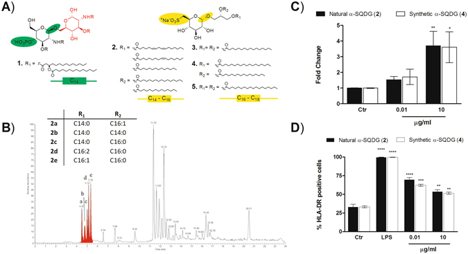
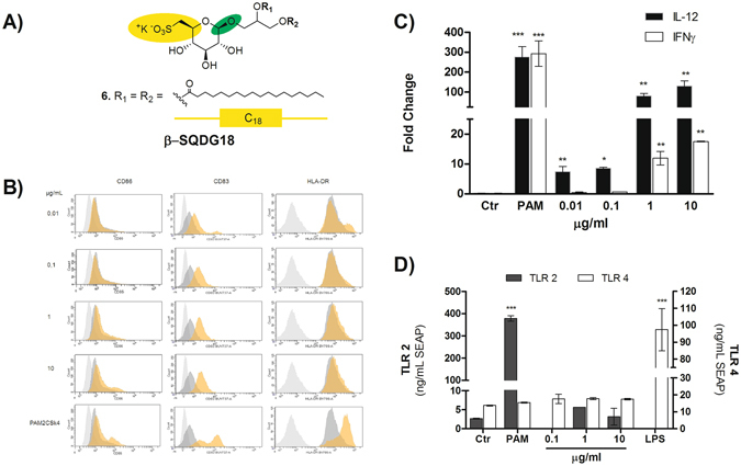
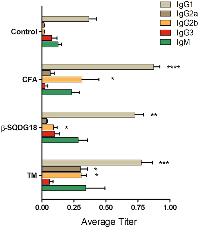
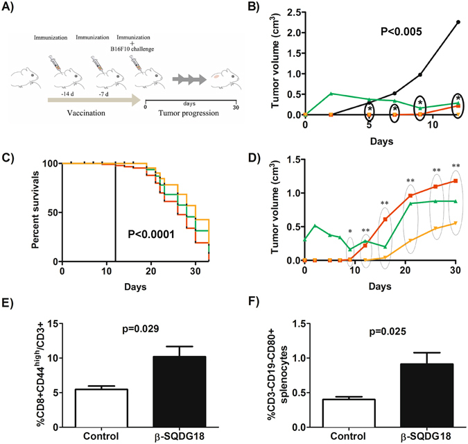
Dendritic Cells (DCs) recognize infectious non-self molecules and engage the adaptive immune system thereby initiating long lasting, antigen-specific responses. As such, the ability to activate DCs is considered a key tool to enhance the efficacy and quality of vaccination. Here we report a novel immunomodulatory sulfolipid named β-SQDG18 that prototypes a class of natural-derived glycolipids able to prime human DCs by a TLR2/TLR4-independent mechanism and trigger an efficient immune response in vivo. β-SQDG18 induces maturation of DC with the upregulation of MHC II molecules and co-stimulatory proteins (CD83, CD86), as well as pro-inflammatory cytokines (IL-12 and INF-γ). Mice immunized with OVA associated to β-SQDG18 (1:500) produced a titer of anti-OVA Ig comparable to traditional adjuvants. In an experimental model of melanoma, vaccination of C57BL/6 mice with β-SQDG18-adjuvanted hgp10 peptide elicited a protective response with a reduction in tumour growth and increase in survival.


Conflict of interest statement

The authors declare that they have no competing interests.

Figures

Figure 1
Figure 1
Chemical structure and DC activation of natural sulfoquinovosides (α-SQDG) from marine diatoms. (A) Structures of DC agonists: monophosphoryl lipid A (MPLA, 1) (reference compound), natural α-SQDG (2) and synthetic (3-5) α-SQDG. Colors underline the chemical differences in type and position of the anionic function, configuration of the anomeric carbon, second sugar residue and fatty acid acyl chain between MPLA (1) and α-SQDG (2-5); (B) LC-MS profile of active α-SQDG (2) in the extract of the marine diatom Thalassiosira weissflogii. The insert reports the type and distribution of fatty acids bound to the sulfoquinovosyl glycerol residue; (C) expression of IL-12 (mean/standard deviation) and (D) expression of HLA-DR by MoDCs stimulated with natural and synthetic analogs of α-SQDG at 10 ng/mL and 10 µg/mL. Results (mean +/− standard deviation) are reported as percentage of positive cells (marker expression value over 104) out of the total DC population. LPS was used as positive control. Asterisks indicate significant differences from the cells treated only with vehicle (control, Ctr) at a 95% (P < 0.05) confidence level, as determined using two-way ANOVA analysis.
Figure 2
Figure 2
Dose-dependent priming of DC by βSQDG18 (6). (A) The chimeric molecule β-SQDG18 (6) derives from combination of the structural characteristics of α-SQDG (yellow) and MPLA (green); (B) Flow-cytometry analysis of the phenotyping markers of DC maturation (HLA-DR, CD83 and CD86) at concentrations between 10 ng/ml to 10 µg/ml of β-SQDG18 (6). gray = isotype control; dark gray = unstimulated cells; orange = stimulated cells. Pam2CSK4 (PAM) was used as positive control; (C) expression of IL-12 and IFN-γ by DC stimulated with β-SQDG18 in the same range of concentrations used for the above marker phenotyping; (D) evaluation of the Toll-Like Receptor (TLR) activation by increasing concentration concentrations of β-SQDG18 in TLR-2 and TLR-4 NF-kB/SEAP reporter cell lines. Pam2CSK4 (PAM) and LPS were used as positive controls for TLR-2 and TLR-4 respectively. Asterisks indicate significant differences from the cells treated only with vehicle (control, Ctr) at a 95% (P < 0.05) confidence level, as determined using two-way ANOVA analysis.
Figure 3
Figure 3
Humoral response of mice after immunization to ovalbumin formulated with β-SQDG18. C56 BL/6 mice were divided into 4 groups and inoculated twice with 5 µg antigen (OVA) in presence or absence (Control) of 2.5 mg β-SQDG18 (1:500 w/w). Co-administrations of OVA together with Complete Freund’s Adjuvant (FCA) or TiterMax (TM) were used as positive controls. Serum anti-OVA antibodies were measured by ELISA and results are shown as endpoint titer (mean/standard error). No increase in OVA-specific IgA was observed. Asterisks indicate significant differences from the control group at a 95% (P < 0.05) confidence level, as determined using two-way ANOVA analysis.
Figure 4
Figure 4
Protective effect of hgp10025–33 peptide formulated with synthetic β-SQDG18 as adjuvant in an experimental model of anti-melanoma vaccine. (A) Experimental design of immunization in C57BL/6 J mice challenged by 105 B16F10 melanoma cells (day 0). The animals were pretreated with the adjuvanted antigen hgp10025–33 peptide (100 μg mouse/injection) on days −14, −7 and 0. For the experiment with β-SQDG18 (600 μg mouse/injection), the antigen was vigorously mixed with a homogeneous suspension of the molecule prior to injection. Mice immunized with the melanoma epitope in association to either CPG oligodeoxynucleotide (30 μg/injection) or Freund’s adjuvants (1:1 vol/vol) were used as positive controls. Unimmunized mice were used as negative control. Tumour volume of melanoma lesions and survival rate were monitored for 30 days after challenging. Each group was composed of eight animals; (B) Tumour growth and (C) percent survival in the four groups of mice. Statistically significant difference between the negative control and each of the three immunized groups of mice is indicated. Black line: negative control; Red line: antigen plus β-SQDG18; Green line: antigen plus Freund’s adjuvant; Orange line: antigen plus CpG; (D) Detail of tumour growth in response to vaccination with the three adjuvants. Groups are indicated by colours as above Asterisks indicate statistically significant differences in comparison to negative control. *P < 0.01; **P < 0.002; (E) Percentage of CD8+CD44high memory T cell in splenocytes of tumour-challenged mice in the negative control group or after vaccination by β-SQDG18 as adjuvant (black column); (F) Percentage of CD3-CD19-CD80+ cells in splenocytes of tumour challenged mice in the negative control group and after vaccination by β-SQDG18 as adjuvant (black column).

References

    1. Wu TY-H, et al. Rational design of small molecules as vaccine adjuvants. Sci. Transl. Med. 2014;6:263ra160. doi: 10.1126/scitranslmed.3009980. - DOI - PubMed
    1. Egli A, et al. Vaccine adjuvants - Understanding molecular mechanisms to improve vaccines. Swiss Med. Wkly. 2014;144:w13940. - PubMed
    1. Coffman RL, Sher A, Seder RA. Vaccine adjuvants: Putting innate immunity to work. Immunity. 2010;33:492–503. doi: 10.1016/j.immuni.2010.10.002. - DOI - PMC - PubMed
    1. Bergmann-Leitner E, Leitner W. Adjuvants in the Driver’s Seat: How Magnitude, Type, Fine Specificity and Longevity of Immune Responses Are Driven by Distinct Classes of Immune Potentiators. Vaccines. 2014;2:252–296. doi: 10.3390/vaccines2020252. - DOI - PMC - PubMed
    1. De Gregorio E, Rappuoli R. From empiricism to rational design: a personal perspective of the evolution of vaccine development. Nat. Rev. Immunol. 2014;14:505–14. doi: 10.1038/nri3694. - DOI - PMC - PubMed

Publication types