. 2017 Jul 24;7(1):6312.
doi: 10.1038/s41598-017-06395-6.

Ryanodine receptors are part of the myospryn complex in cardiac muscle

Affiliations

Ryanodine receptors are part of the myospryn complex in cardiac muscle

Matthew A Benson et al. Sci Rep. .

Abstract

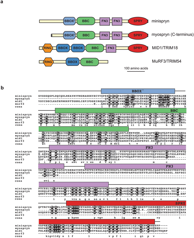
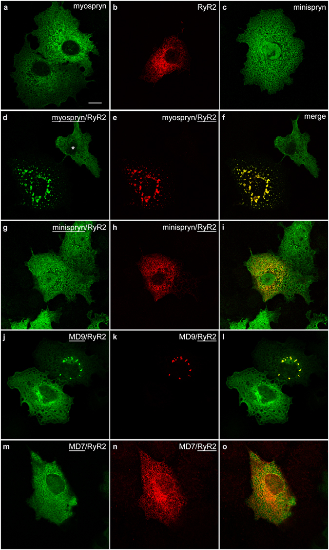
The Cardiomyopathy-associated gene 5 (Cmya5) encodes myospryn, a large tripartite motif (TRIM)-related protein found predominantly in cardiac and skeletal muscle. Cmya5 is an expression biomarker for a number of diseases affecting striated muscle and may also be a schizophrenia risk gene. To further understand the function of myospryn in striated muscle, we searched for additional myospryn paralogs. Here we identify a novel muscle-expressed TRIM-related protein minispryn, encoded by Fsd2, that has extensive sequence similarity with the C-terminus of myospryn. Cmya5 and Fsd2 appear to have originated by a chromosomal duplication and are found within evolutionarily-conserved gene clusters on different chromosomes. Using immunoaffinity purification and mass spectrometry we show that minispryn co-purifies with myospryn and the major cardiac ryanodine receptor (RyR2) from heart. Accordingly, myospryn, minispryn and RyR2 co-localise at the junctional sarcoplasmic reticulum of isolated cardiomyocytes. Myospryn redistributes RyR2 into clusters when co-expressed in heterologous cells whereas minispryn lacks this activity. Together these data suggest a novel role for the myospryn complex in the assembly of ryanodine receptor clusters in striated muscle.


Conflict of interest statement

The authors declare that they have no competing interests.

Figures

Figure 1
Figure 1
Minispryn bioinformatics. (a) Domain architecture of minispryn and related proteins. Minispryn, myospryn and two members of the TRIM-protein family share extensive sequence similarity at their C-termini. While the TRIM proteins are E3 ubiquitin ligases, the absence of the RING domain in minispryn and myospryn probably preclude this function. (b) Multiple sequence alignment. The complete coding sequence of minispryn aligns with amino acids 3171–3739 of myospryn (CAE02649.1), 139–706 of midline-1 (XP_017173902) and 123–366 of murf3 (NP_067422.1) spanning the BBOX/BBOX’ and BBC domains of each protein and the FN3 and SPRY domains of myospryn and midline-1.
Figure 2
Figure 2
Minispryn transcript and protein. (a) Northern blot showing the size and tissue distribution of the murine minispryn transcript. A single band of approximately 7 kb is only detected in heart and skeletal muscle. The blot was stripped and re-hybridized with a GAPDH probe to demonstrate the presence of mRNA in each lane. Key: B, brain; H, heart; Lu, lung; Li, liver; Ki, kidney; St, stomach; SI, small intestine; M, muscle; O, ovary; Th, thymus; Te, testes; U, uterus; PU, pregnant uterus; Pl, placenta; Sp, spleen. (b) Western blot of several mouse tissues showing that minispryn is a protein of approximately 90 kDa that is found predominantly in heart and skeletal muscle. (c) Antibody specificities were determined in heterologous cells. Lysates prepared from HEK-293T cells expressing full-length myospryn (clone 21B) and minispryn (clone 7E) were processed for immunoblotting with antibodies detecting their cognate antigen. The 819 antibody only detects minispryn in transfected cells whereas des122 only detects myospryn under the same conditions. The lysates were also immunoblotted with anti-tubulin antibodies to demonstrate similar levels of protein in each transfection. Lysates prepared from untransfected HEK-293T cells are also shown. (d) In tissue, minispryn and myospryn were immunoprecipitated from RIPA extracts of cardiac muscle using specific antibodies. Minispryn and myospryn are robustly immunoprecipitated with their cognate antisera but also co-IP with the antisera raised against the paralogous protein. Neither myospryn nor minispryn are immunoprecipitated with unrelated IgG. (e) Minispryn and myospryn similarly co-immunoprecipitate from RIPA extracts of skeletal muscle. X denotes an empty lane.
Figure 3
Figure 3
Immunoaffinity purification of the myospryn complex from cardiac muscle. (a) Proteins eluted from an anti-minispryn immunoaffinity column or protein G only column were separated by PAGE and stained with colloidal Coomassie blue dye. Bands A1-A3 that were only present in minispryn immunoaffinity eluate were excised and processed for mass spectrometry. The identity of the major protein(s) found in each band is listed. Note that in this study we focussed exclusively on proteins in bands A1, A2 and A3. Corresponding peptides for myospryn, RyR2 and minispryn are shown in Supplementary Dataset 1. (b) Robust minispryn enrichment following immunoaffinity purification was demonstrated by immunoblotting using the 819 anti-minispryn antibody. The sizes of the molecular weight markers are shown in kDa.
Figure 4
Figure 4
Immunolocalization of myospryn and minispryn in isolated cardiomyocytes. Enzymatically isolated guinea pig ventricular cardiomyocytes were fixed and processed for confocal immunofluorescence microscopy using the indicated antibodies. (ac) At low magnification myospryn (a) immunoreactivity is visible in discontinuous striations that run perpendicular to the long axis of the cardiomyocyte partially overlapping the nucleus (arrow in a). (df) Higher magnification images show that myospryn immunoreactivity is present in distinct punctae that co-localise with ryanodine receptors (e) in the SR (f and inset). (gi) Anti-minispryn (g) immunoreactivity also appears in discontinuous striations running the length of the cardiomyocyte and is also present in the nucleus (arrow in g). (jl) In common with myospryn, higher magnification images show that minispryn partially co-localises with ryanodine receptors (k) in the SR (l and inset). (m–o) Myospryn immunoreactivity overlaps the Z-line labelled with an anti-alpha-actinin antibody (m, n, o). (pr) Similarly, minispryn immunoreactivity also partially overlaps alpha-actinin at the Z-line (p, q, r). (sx) Cardiomyocytes labelled with either anti-SERCA2 (s, t, u) or anti-sarcalumenin (v, w, x) antibodies and myospryn antibodies demonstrate that myospryn immunoreactivity (arrowheads in s) appears to be restricted to the j-SR rather than the l-SR (arrows in t).
Figure 5
Figure 5
Myospryn clusters RyR2 in heterologous cells. COS-7 cells were transfected with expression constructs as indicated. (ac) Full-length myospryn (a) and minispryn (c) appear to be associated with the internal membranes of the cell when expressed on their own whereas RyR2 (b) labelling is restricted to the endoplasmic reticulum. (df) Co-expression of myospryn (d) and RyR2 (e) results in the clustering of both proteins in intensely staining punctae (merged image, f). Note that myospryn is not clustered in the adjacent cell (asterisk) that does express RyR2. (gi) By contrast co-expression of minispryn (g) and RyR2 (h) does not alter the distribution of either protein (merged image, i). (j o) Truncated myospryn constructs were used to delineate the regions of the protein involved in RyR2 clustering. MD9 (j) was able to cluster RyR2 (k and merged image, l) whereas the smaller protein MD7 (m) failed to cluster the receptor (n and merged image, o). The underlining denotes the transfected construct for each channel. Scale bar = 10 µm.

References

    1. Clark KA, McElhinny AS, Beckerle MC, Gregorio CC. Striated muscle cytoarchitecture: an intricate web of form and function. Annu Rev Cell Dev Biol. 2002;18:637–706. doi: 10.1146/annurev.cellbio.18.012502.105840. - DOI - PubMed
    1. Frank D, Kuhn C, Katus HA, Frey N. The sarcomeric Z-disc: a nodal point in signalling and disease. J Mol Med (Berl) 2006;84:446–468. doi: 10.1007/s00109-005-0033-1. - DOI - PubMed
    1. Lange S, Ehler E, Gautel M. From A to Z and back? Multicompartment proteins in the sarcomere. Trends Cell Biol. 2006;16:11–18. doi: 10.1016/j.tcb.2005.11.007. - DOI - PubMed
    1. Kontrogianni-Konstantopoulos A, Bloch RJ. Obscurin: a multitasking muscle giant. J Muscle Res Cell Motil. 2005;26:419–426. doi: 10.1007/s10974-005-9024-7. - DOI - PubMed
    1. McElhinny AS, Kazmierski ST, Labeit S, Gregorio CC. Nebulin: the nebulous, multifunctional giant of striated muscle. Trends Cardiovasc Med. 2003;13:195–201. doi: 10.1016/S1050-1738(03)00076-8. - DOI - PubMed

Publication types

Substances