. 2017 Jul 24;7(1):6215.
doi: 10.1038/s41598-017-06353-2.

Fibulin-3 promotes osteosarcoma invasion and metastasis by inducing epithelial to mesenchymal transition and activating the Wnt/β-catenin signaling pathway

Affiliations

Fibulin-3 promotes osteosarcoma invasion and metastasis by inducing epithelial to mesenchymal transition and activating the Wnt/β-catenin signaling pathway

Songgang Wang et al. Sci Rep. .

Retraction in

Abstract

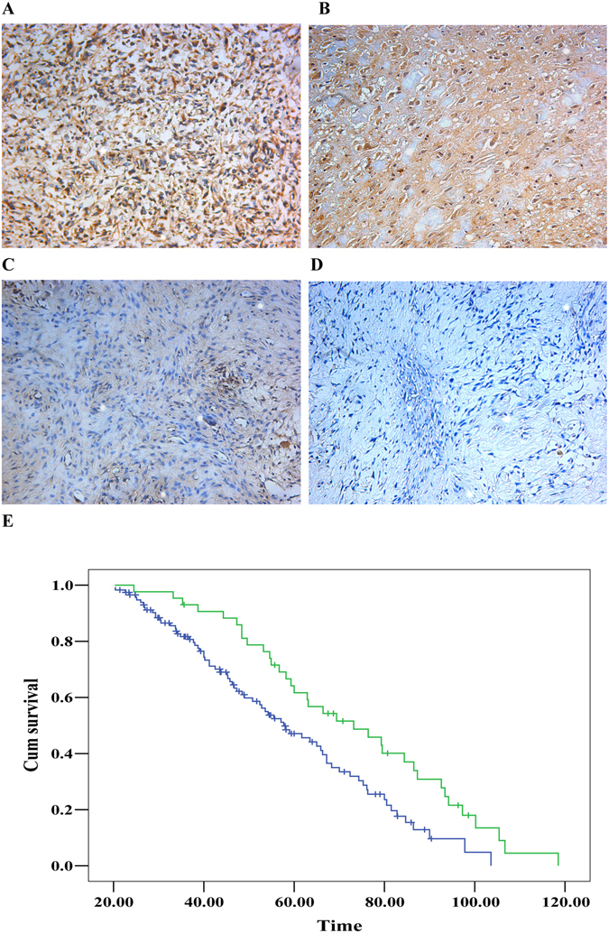
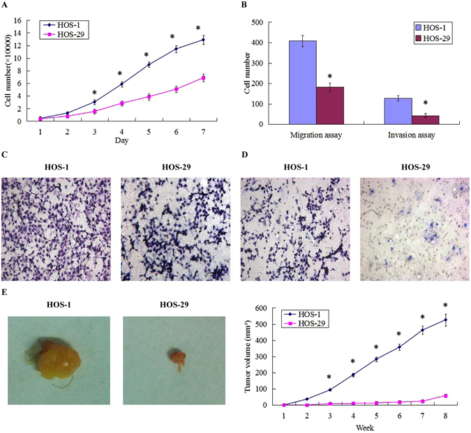
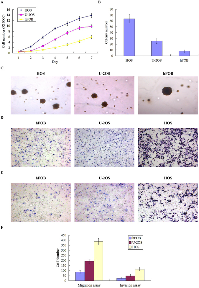
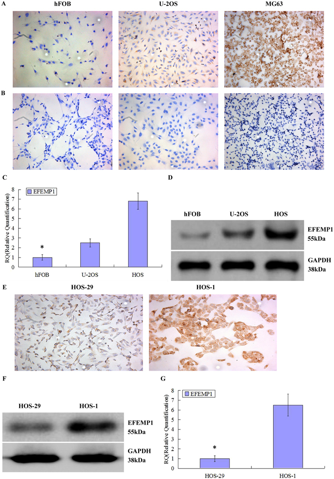
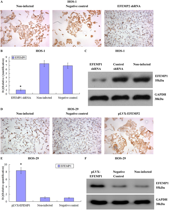
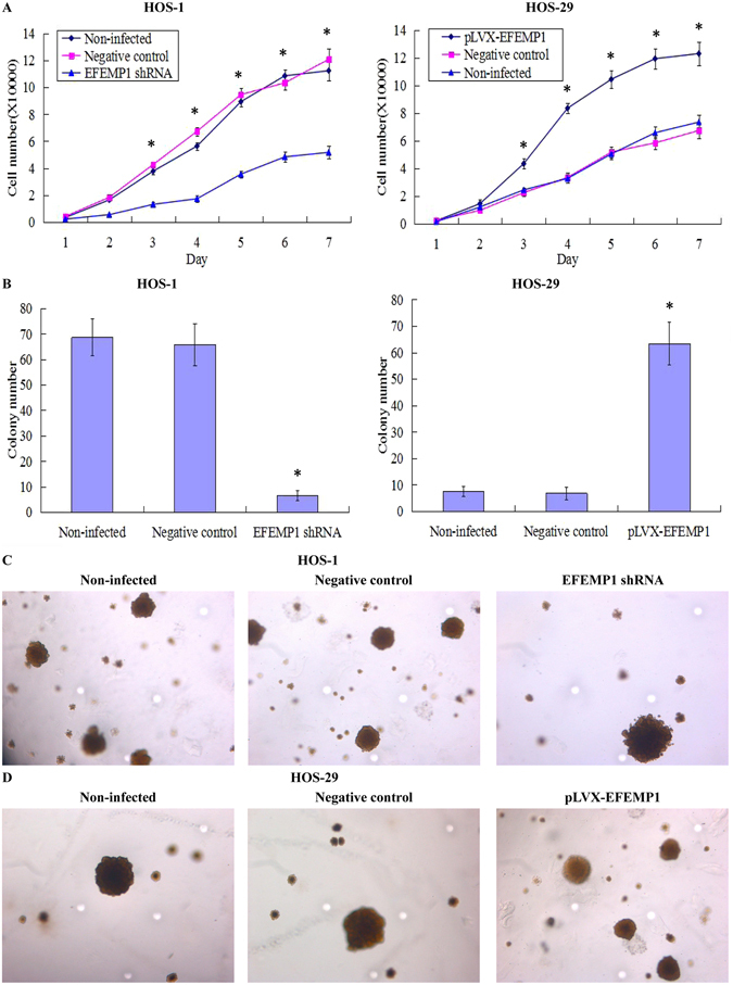
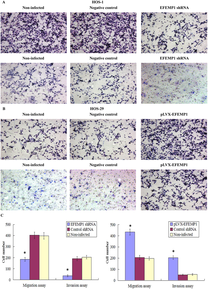
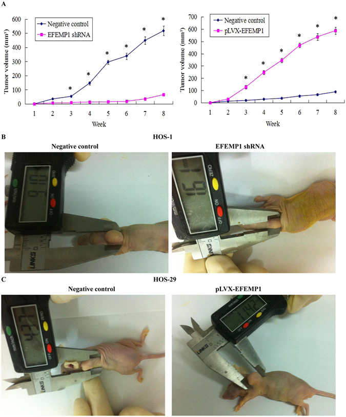
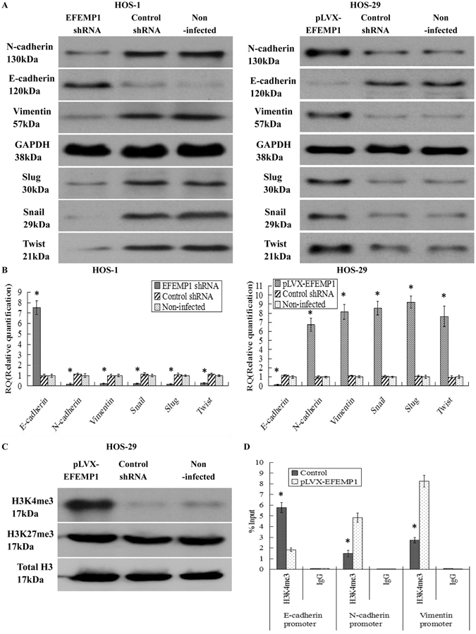
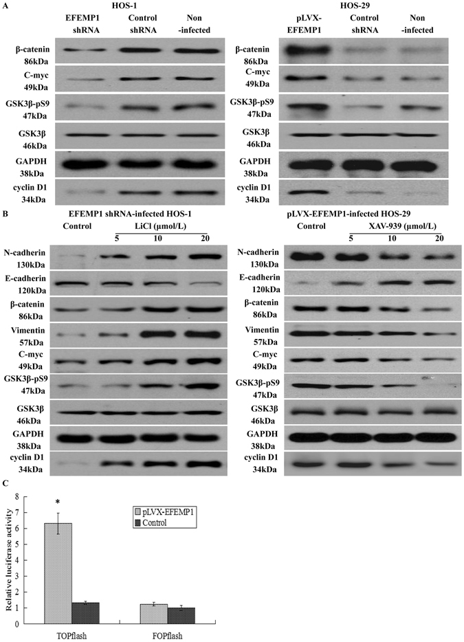
This study explored the role of fibulin-3 in osteosarcoma progression and the possible signaling pathway involved. Fibulin-3 mRNA and protein expression in normal tissue, benign fibrous dysplasia, osteosarcoma, osteosarcoma cell lines (HOS and U-2OS), the normal osteoblastic cell line hFOB, and different invasive subclones was evaluated by immunohistochemistry (IHC) or immunocytochemistry (ICC) and real time reverse transcriptase-polymerase chain reaction (real time qRT-PCR). To assess the role of fibulin-3 in the invasion and metastasis of osteosarcoma cells, lentiviral vectors with fibulin-3 small hairpin RNA (shRNA) and pLVX-fibulin-3 were constructed and used to infect the highly invasive and low invasive subclones. The effects of fibulin-3 knockdown and upregulation on the biological behavior of osteosarcoma cells were investigated by functional in vitro and in vivo assays. The results revealed that fibulin-3 expression was upregulated in osteosarcoma, and was positively correlated with low differentiation, lymph node metastasis, and poor prognosis. Fibulin-3 could promote osteosarcoma cell invasion and metastasis by inducing EMT and activating the Wnt/β-catenin signaling pathway. Collectively, our findings demonstrate that fibulin-3 is a promoter of osteosarcoma development and progression, and suggest a novel therapeutic target for future studies.


Conflict of interest statement

The authors declare that they have no competing interests.

Figures

Figure 1
Figure 1
Expressions of fibulin-3 in human osteosarcoma tissues. Fibulin-3 expressions in osteosarcoma tissue (A,B), fibrous dysplasia tissue (C) and normal tissue (D) were measured by IHC. (E) Patients with high fibulin-3 expression (blue line) had a much worse prognosis than those with low fibulin-3 expression (green line). (Magnification ×200).
Figure 2
Figure 2
Establishment of high invasive subclone and low invasive subclone. (A) The high invasive subclone had the higher proliferative abilities than the low invasive subclone. (B) The average counts of migrating and invading cells of high invasive subclone were both much higher than those of low invasive subclone. (C) The images of cells crossing PVPF filters without Matrigel were examined by cell migration assay. (D) The images of cells crossing PVPF filters with Matrigel were examined by cell invasion assay. (E) The tumor volumes formed by high invasive subclone were much larger than those formed by low invasive subclone after subcutaneous inoculation. (F) Tumor growths of high invasive subclone and low invasive subclone were observed continuously for 2 months. (Magnification ×200). *P < 0.05.
Figure 3
Figure 3
Different proliferation, migration and invasion abilities of human osteosarcoma cell lines and normal osteoblastic cell line. (A) The human osteosarcoma cell lines U-2OS and HOS had stronger proliferative abilities than normal osteoblastic cell line hFOB. (B) The number of colonies formed by HOS and U-2OS was also significantly greater than that formed by hFOB. (C) The colony images of human osteosarcoma cell lines HOS and U-2OS and normal osteoblastic cell line hFOB were examined by soft agar colony formation assay. (D) The images of cells migrating PVPF filters were examined by cell migration assay using Boyden chambers. (E) The images of cells invading Matrigel-coated membranes were examined by cell invasion assay using Boyden chambers. (F) The average counts of migrating and invading cells of human osteosarcoma cell lines HOS and U-2OS were both much higher than those of normal osteoblastic cell line hFOB. (Magnification ×200). *P < 0.05.
Figure 4
Figure 4
Fibulin-3 expressions in human osteosarcoma cell lines and in differently invasive subclones. fibulin-3 expressions in human osteosarcoma cell lines HOS and U-2OS and normal osteoblastic cell line hFOB were measured by ICC staining. (A) Brown granules were detected in the cell cytoplasm. (B) Negative controls were obtained by replacing the primary antibody with PBS. (C) Real time q-RT-PCR and (D) western blot were performed to detect the mRNA and protein expressions of fibulin-3 in human osteosarcoma cell lines and normal osteoblastic cell line. Fibulin-3 expressions in high invasive subclone and low invasive subclone were measured by (E) ICC staining, (F) western blot (cropped blot) (see full blots in the supplementary information) and (G) q-RT-PCR. (Magnification ×200). *P < 0.05.
Figure 5
Figure 5
Identification of downregulated and upregulated fibulin-3 expression in lentivirus transfection systems. Fibulin-3 expressions in non-infected cells, control shRNA infected cells and fibulin-3 shRNA infected cells were measured by (A) ICC staining, (B) real time q-RT-PCR and (C) Western blot. Fibulin-3 expressions in non-infected cells, control shRNA infected cells and pLVX-fibulin-3 infected cells were measured by (D) ICC staining, (E) real time q-RT-PCR and (F) Western blot (cropped blot). (Magnification ×200). *P < 0.05.
Figure 6
Figure 6
Effect of fibulin-3 knockdown and overexpression on osteosarcoma cell proliferation and colony formation abilities. (A) Downregulated fibulin-3 markedly inhibited cell proliferation of the highly invasive subclone HOS-1, whereas upregulated fibulin-3 significantly promoted cell proliferation of the low invasive subclone HOS-29. (B) The colony forming efficiencies of fibulin-3-silenced cells were decreased, and conversely, upregulation of fibulin-3 could increase the colony forming efficiency of the low invasive subclone. (C) The colony images of non-infected cells, control shRNA infected cells and fibulin-3 shRNA infected cells were examined by soft agar colony formation assay. (D) The colony images of non-infected cells, control shRNA infected cells and pLVX-fibulin-3 infected cells were examined by soft agar colony formation assay. (Magnification × 200). *P < 0.05.
Figure 7
Figure 7
Effect of fibulin-3 knockdown and overexpression on osteosarcoma cell migration and invasion. (A) Cell migration and invasion images of non-infected cells, control shRNA infected cells and fibulin-3 shRNA infected cells were measured by cell migration and invasion assays using Boyden chambers. (B) Cell migration and invasion images of non-infected cells, control shRNA infected cells and pLVX-fibulin-3 infected cells were measured by cell migration and invasion assays using Boyden chambers. (C) The average counts of migrating and invading fibulin-3 shRNA infected cells were much lower than those of the negative controls and non-infected groups; meanwhile, the average counts of migrating and invading pLVX-fibulin-3 infected cells were much higher than those of the negative controls and the non-infected groups. (Magnification × 200). *P < 0.05.
Figure 8
Figure 8
Effects of fibulin-3 knockdown and overexpression on tumor growth in a xenograft model. (A) Tumor growths in lentivirus transfection systems were observed continuously for 8 weeks. (B) The average volumes of the tumors formed in the fibulin-3 shRNA infected group were much lower than those formed by the negative control HOS-1. (C) The average volumes of the tumors formed by pLVX-fibulin-3 infected cells were much higher than those formed by the negative control HOS-29. *P < 0.05.
Figure 9
Figure 9
Effects of fibulin-3 knockdown and overexpression on EMT genes correlated to tumor progression. After lentivirus transfections, EMT markers, including E-cadherin, N-cadherin, vimentin, Snail Slug and Twist were measured by (A) Western blot (cropped blot) and (B) real-time q-RT-PCR in the lentivirus transfection systems. (C) H3K4me3 and H3K27me3 expressions were assessed by Western blot. Histone H3 was used as a loading control. H3K4me3 is associated with transcriptional activation, and H3K27me3 was responsible for turning off transcription. Ectopic expression of fibulin-3 only increased H3K4me3 modification but had no effect on H3K27me3 modification. (D) Quantitative chromatin immunoprecipitation was performed to assess H3K4me3 occupancy in pLVX-EFEMP1 cells. IgG was used as the negative control. “% input” indicated the ratio of DNA fragment of each promoter region bound by H3K4me3 to the total amount of input DNA fragment without H3K4me3 antibody pull-down. *P < 0.05.
Figure 10
Figure 10
Effects of fibulin-3 on the Wnt/β-catenin signaling pathway by Western blot (cropped blot). (A) Fibulin-3 knockdown reduced the expression of GSK3β-pS9, β-catenin, C-myc, and cyclin D1; in contrast, fibulin-3 upregulation increased the expression of GSK3β-pS9, β-catenin, C-myc, and cyclin D1. (B) Wnt signaling pathway activator LiCl could promote the Wnt/β-catenin pathway and EMT, both of which were deactivated by fibulin-3 knockdown; meanwhile, Wnt signaling pathway inhibitor XAV-939 could significantly inhibit the Wnt/β-catenin pathway and EMT, both of which were activated by fibulin-4 upregulation. (C) Cells were transfected with TOPFLASH or FOPFLASH construct. After 48 h, luciferase activity was determined by the dual-luciferase system. The pRL-TK Renilla luciferase reporter construct was co-transfected in each sample to normalize transfection efficiency. The luciferase activity of the reporter is expressed relative to the activity in control cells, which is defined as 1.0. *P < 0.05.

Similar articles

Cited by

References

    1. Geller DS, Gorlick R. Osteosarcoma: a review of diagnosis, management, and treatment strategies. Clin Adv Hematol Oncol. 2010;8(10):705–718. - PubMed
    1. Messerschmitt PJ, Rettew AN, Brookover RE, et al. Specific tyrosine kinase inhibitors regulate human osteosarcoma cells in vitro. Clin Orthop Relat Res. 2008;466(9):2168–2175. doi: 10.1007/s11999-008-0338-9. - DOI - PMC - PubMed
    1. Messerschmitt PJ, Garcia RM, Abdul-Karim FW, Greenfield EM, Getty PJ. Osteosarcoma. J Am Acad Orthop Surg. 2009;17:515–527. doi: 10.5435/00124635-200908000-00005. - DOI - PubMed
    1. Zhang Y, Marmorstein LY. Focus on molecules: fibulin-3 (EFEMP1) Exp Eye Res. 2010;90(3):374–5. doi: 10.1016/j.exer.2009.09.018. - DOI - PMC - PubMed
    1. De Vega S, Iwamoto T, Yamada Y. Fibulins: multiple roles in matrix structures and tissue functions. Cell Mol Life Sci. 2009;66:1890–1902. doi: 10.1007/s00018-009-8632-6. - DOI - PMC - PubMed

Publication types

MeSH terms