. 2017 Jul 24;7(1):6238.
doi: 10.1038/s41598-017-06217-9.

A modified formulation of Huanglian-Jie-Du-Tang reduces memory impairments and β-amyloid plaques in a triple transgenic mouse model of Alzheimer's disease

Affiliations

A modified formulation of Huanglian-Jie-Du-Tang reduces memory impairments and β-amyloid plaques in a triple transgenic mouse model of Alzheimer's disease

Siva Sundara Kumar Durairajan et al. Sci Rep. .

Abstract

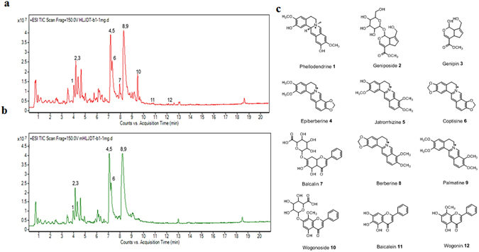
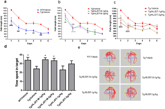
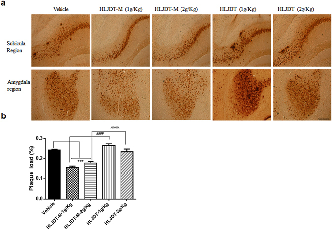
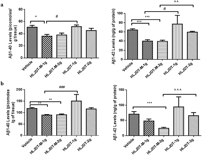
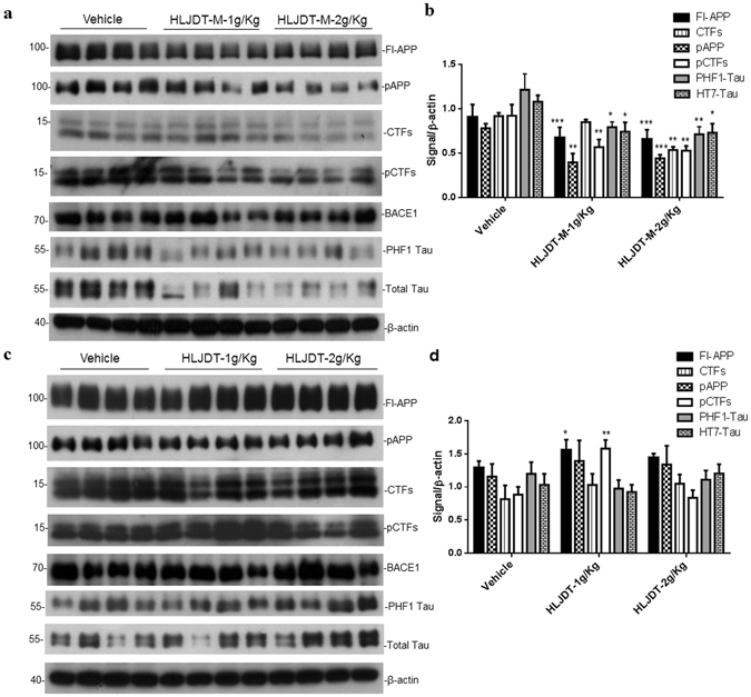
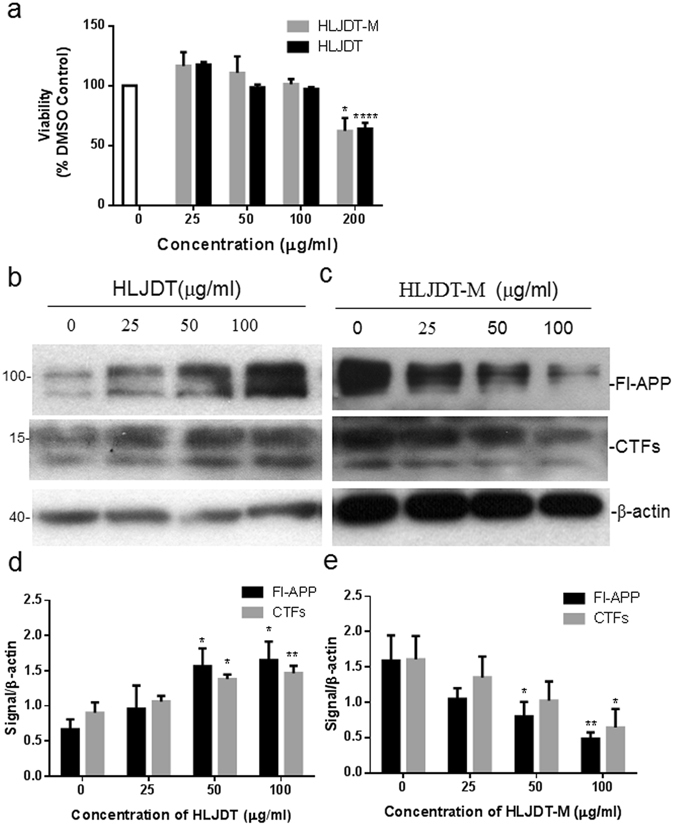
Alzheimer's disease (AD) is a degenerative disorder typified by progressive deterioration of memory and the appearance of β-amyloid peptide (Aβ)-rich senile plaques. Recently we have identified a novel function of a patented formulation of modified Huanglian-Jie-Tu-Tang (HLJDT-M), a Chinese herbal medicine, in treating AD in in vitro studies (US patent No. 9,375,457). HLJDT-M is a formulation composed of Rhizoma Coptitis, Cortex Phellodendri and Fructus Gardeniae without Radix Scutellariae. Here, we assessed the efficacy of HLJDT-M on a triple transgenic mouse model of AD (3XTg-AD). Oral administration of HLJDT-M ameliorated the cognitive dysfunction of 3XTg-AD mice and lessened the plaque burden. In addition, biochemical assays revealed a significant decrease in levels of detergent-soluble and acid-soluble Aβ via decreasing the levels of full length amyloid-β precursor protein (FL-APP) and C-terminal fragments of APP (CTFs) in brain lysates of HLJDT-M-treated mice. HLJDT-M treatment also significantly reduced the levels of FL-APP and CTFs in N2a/SweAPP cells. In contrast, treatment using the classical formula HLJDT did not reduce the memory impairment of 3XTg-AD mice and, rather, increased the Aβ/Fl-APP/CTFs in both animal and cell culture studies. Altogether, our study indicates that HLJDT-M is a promising herbal formulation to prevent and/or cure AD.


Conflict of interest statement

The authors declare that they have no competing interests.

Figures

Figure 1
Figure 1
LC-ESI-Q/TOF chromatograms (TIC) of HLJDT (a) and HLJDT-M (b). (c). Chemical structure of representative compounds of HLJDT. Peaks: 1. Phellodendrine, 2. Geniposide, 3. Genipin, 4. Epiberberine, 5. Jaterorhizine, 6. Coptisine, 7. Baicalin, 8. Berberine, 9. Palmatine, 10. Wogonoside, 11. Baicalein, 12. Wogonin.
Figure 2
Figure 2
Behaviour study of HLJDT and HLJDT-M on 3XTg-AD mice. Acquisition of spatial memory was evaluated by Morris water maze (hidden platform) in HLJDT, HLJDT-M and vehicle-treated 3XTg-AD mice (a,b,c). Results are represented as the mean length values ± standard error of the mean (SEM) of all mice from 4 trials per day (n = 10). In probe trial, mice are evaluated for the amount of time spent in searching the platform location after 24-hour retention trial in MWM (d,e). In the probe trial, the HLJDT-M treated mice stayed longer in the target quadrant than the vehicle-treated mice, showing memory retention. The symbol denotes statistical differences among the given groups over all trial days. ###p < 0.001 (WT treated with vehicle vs. Tg treated with vehicle); ***p < 0.001 (Tg treated with HLJDT-M at 1 g/kg vs. Tg treated with vehicle), ~~~p < 0.001 (Tg treated with 2 g/kg of HLJDT-M vs. Tg treated with vehicle), ^^p < 0.01 (Tg treated with 1 g/kg of HLJDT vs. Tg treated with vehicle) and ΦΦΦp < 0.001 (Tg treated with 2 g/kg of HLJDT vs. Tg treated with vehicle).
Figure 3
Figure 3
HLJDT-M, but not HLJDT, reduces hippocampal Aβ-plaque pathology in 3XTg-AD mice. Immunohitochemical labelling of Aβ with 4G8 antibody in representative figures taken from the coronal sections in 3XTg-AD mice were orally administered with HLJDT-M or HLJDT at doses of 1 and 2 g/kg per day for 6 months.
Figure 4
Figure 4
HLJDT-M but not HLJDT treatment reduces Aβ peptide levels in 3XTg-AD mice. SDS-soluble (a) and formic acid-soluble (b) Aβ1-40 and Aβ1-42 levels from the right brain hemisphere were measured by sandwich ELISA. Both Aβ1-40 and Aβ1-42 were reduced in the brains lysates of HLJDT-M-treated (**p < 0.01) animals. Compared to the HLJDT-M treatment groups, HLJDT significantly increased both detergent and acid-soluble Aβ1-40 and Aβ1-42 levels. Values denote group mean ± SEM. The statistical significant difference between the groups are denoted as *p < 0.05, **p < 0.01, ***p < 0.001 vs. vehicle control; #p < 0.05, ###p < 0.001 for 1 g/kg HLJDT-M vs. 1 g/kg HLJDT and ^p < 0.05, ^^^p < 0.001 for 1 g/kg HLJDT-M vs. 1 g/kg HLJDT. N = 10 mice per group.
Figure 5
Figure 5
Different treatment effects of HLJDT-M (a) and HLJDT (a) on the levels of APP metabolites, phospho-Tau and total Tau in 3XTg-AD mice. Immunoblot presenting the levels of FL-APP, CTFs (CTFα and CTFβ), pAPP and pCTFs (α and β). Quantification of immunoblots by densitometric analysis and is presented as the ratio of FL-APP, CTFs, pAPP, PHF-1 Tau, total Tau and pCTFs against β-actin in the SDS brain lysates of 3XTgAD mice treated with HLJDT-M or HLJDT or vehicle (c and d). The statistical significance are denoted as *p < 0.05, **p < 0.01, **p < 0.001 when compared with vehicle-treated 3XTg-AD mice. Data represent mean ± SEM. N = 8 mice per group.
Figure 6
Figure 6
Treatment of N2a-SwedAPP cells with HLJDT-M and HLJDT differentially modulated the processing of APP. Cells incubated with aqueous extract of HLJDT or HLJDT-M at three different concentrations (25, 50 or 100 μg/ml) or with vehicle for 48 hours. After incubation, cell lysates were probed for APP and CTFs by Western blot. The statistical significance among different groups are represented as *p < 0.05, **p < 0.01, **p < 0.001.

References

    1. Selkoe DJ. Cell biology of protein misfolding: the examples of Alzheimer’s and Parkinson’s diseases. Nat Cell Biol. 2004;6:1054–1061. doi: 10.1038/ncb1104-1054. - DOI - PubMed
    1. Ittner LM, Gotz J. Amyloid-beta and tau–a toxic pas de deux in Alzheimer’s disease. Nat Rev Neurosci. 2011;12:65–72. doi: 10.1038/nrn2967. - DOI - PubMed
    1. Prince, M. et al. Recent global trends in the prevalence and incidence of dementia, and survival with dementia. Alzheimers Res Ther8 (2016). - PMC - PubMed
    1. Cummings J, et al. Drug development in Alzheimer’s disease: the path to 2025. Alzheimers Res Ther. 2016;8:39. doi: 10.1186/s13195-016-0207-9. - DOI - PMC - PubMed
    1. Iqbal K, Grundke-Iqbal I. Alzheimer’s disease, a multifactorial disorder seeking multitherapies. Alzheimers Dement. 2010;6:420–424. doi: 10.1016/j.jalz.2010.04.006. - DOI - PMC - PubMed

Publication types

MeSH terms