. 2017 Jul 24;8(1):102.
doi: 10.1038/s41467-017-00085-7.

p62/SQSTM1/Sequestosome-1 is an N-recognin of the N-end rule pathway which modulates autophagosome biogenesis

Affiliations

p62/SQSTM1/Sequestosome-1 is an N-recognin of the N-end rule pathway which modulates autophagosome biogenesis

Hyunjoo Cha-Molstad et al. Nat Commun. .

Abstract

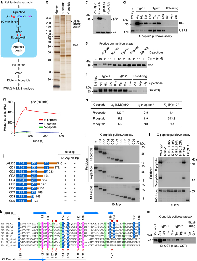
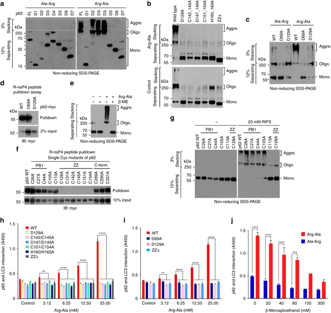
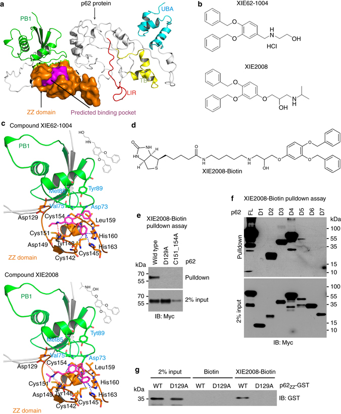
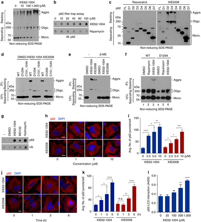
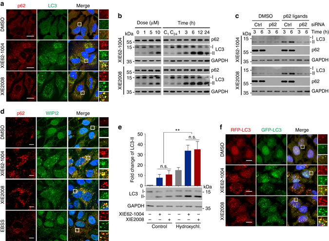
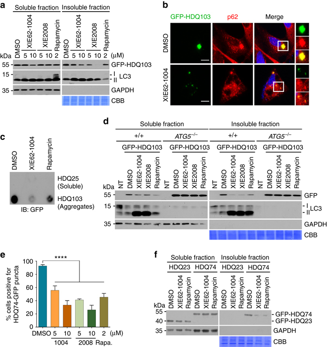
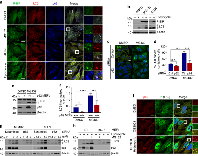
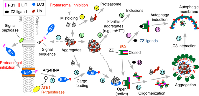
Macroautophagy mediates the selective degradation of proteins and non-proteinaceous cellular constituents. Here, we show that the N-end rule pathway modulates macroautophagy. In this mechanism, the autophagic adapter p62/SQSTM1/Sequestosome-1 is an N-recognin that binds type-1 and type-2 N-terminal degrons (N-degrons), including arginine (Nt-Arg). Both types of N-degrons bind its ZZ domain. By employing three-dimensional modeling, we developed synthetic ligands to p62 ZZ domain. The binding of Nt-Arg and synthetic ligands to ZZ domain facilitates disulfide bond-linked aggregation of p62 and p62 interaction with LC3, leading to the delivery of p62 and its cargoes to the autophagosome. Upon binding to its ligand, p62 acts as a modulator of macroautophagy, inducing autophagosome biogenesis. Through these dual functions, cells can activate p62 and induce selective autophagy upon the accumulation of autophagic cargoes. We also propose that p62 mediates the crosstalk between the ubiquitin-proteasome system and autophagy through its binding Nt-Arg and other N-degrons.Soluble misfolded proteins that fail to be degraded by the ubiquitin proteasome system (UPS) are redirected to autophagy via specific adaptors, such as p62. Here the authors show that p62 recognises N-degrons in these proteins, acting as a N-recognin from the proteolytic N-end rule pathway, and targets these cargos to autophagosomal degradation.


Conflict of interest statement

X.-Q.X. is a consultant for Oxis Biotech. The remaining authors declare no competing financial interests.

Figures

Fig. 1
Fig. 1
Proteomic identification of p62 as an N-recognin. a Schematic representation illustrating the affinity-based proteomic identification of proteins that bind to N-terminal destabilizing residues of synthetic peptides. N-terminal residues of bead-conjugated peptide are indicated by three-letter abbreviations. Biotinylated 11-mer peptides were covalently linked to streptavidine agarose beads. The pulled-down proteins were identified by iTRAQ-MS/MS analysis. b Silver staining of testicular proteins immobilized by 11-mer X-peptides. c, d In vitro peptide pulldown assays of endogenous p62 and UBR2 using HEK293 cells and X-peptides indicated in the figures. e Peptide competition assay using in vitro transcribed and translated p62. f X-peptide pulldown assay using p62-D3 (#1–131) ectopically expressed in HEK293 cells. g Biacore sensorgrams showing the kinetic analysis of p62-D3 binding to Arg-peptide, Phe-peptide or Val-peptide. h Surface plasmon resonance assays measuring the binding of p62 (D3) to X-peptides. Association rate constants k a, dissociation rate constants k d, equilibrium constants K D = k a/k d. i Schematic representation of C-terminal deletion mutants of p62-D3 construct. j X-peptide pulldown assay with constructs shown in i where X is either Arg or Trp. k Alignment of the UBR boxes of UBR1 and UBR2 in comparison with p62 ZZ domain. C2H2 zinc fingers are highlighted in pink, and atypical binuclear zinc fingers are highlighted in blue. Identical residues are highlighted in green, and red circles mark residues that are essential for the recognition of destabilizing N-terminal residues. Residues of the ZZ domain that are mutated to alanine are indicated by the letter A (red). l The inhibitory effect of ZZ point mutations on p62 binding to X-peptide (X=R or W). X-peptide pulldown assay with ZZ point mutants shown in k. m X-peptide pulldown assay using 93-residue p62ZZ-GST containing intact ZZ domain
Fig. 2
Fig. 2
The binding of Nt-Arg to p62 ZZ domain facilitates disulfide bond-linked aggregation of p62 and p62–LC3 interaction. a In vitro oligomerization assay using myc/His tagged p62 deletion mutants (Supplementary Fig. 2a), followed by non-reducing SDS-PAGE and immunoblotting using a mixture of antibodies to p62 and Myc. b In vitro oligomerization assay using myc/His tagged wild type p62 and mutants carrying point mutations in ZZ domain. c In vitro p62 oligomerization assay using p62 D69A and D129A mutants in comparison with wild-type p62. d R-nsP4 pulldown assay using constructs used in c. e In vitro oligomerization assay of p62 ectopically expressed in HEK293 cells treated with 50 mM Arg-Ala in the presence of 50 mM β-mercaptoethanol (β-ME). f R-nsP4 peptide binding assay of p62 Cys mutants. Each Cys/Ala p62 mutant with myc/His tag was expressed in HEK293 cells, and 50 μg of total protein was used for pulldown. g In vitro oligomerization assay of p62 Cys mutants used in f. Cell lysates with ectopically expressed wild-type p62 and Cys mutants that can bind Nt-Arg were incubated with 20 mM RIFS tetrapeptide for 1.5 h at room temperature. h ELISA measuring the interaction of p62 ZZ domain mutants with LC3. Cell lysates overexpressing p62 proteins were incubated with Arg-Ala at different concentrations to allow p62 binding to LC3-GST linked to glutathione coated plates. Shown are the means (±S.D.) of three independent experiments, each performed in triplicate. A one-way ANOVA was performed to determine statistical significance (**P < 0.01; ****P < 0.0001). i ELISA measuring p62D69A interaction with LC3 as described in h. The graph represents the mean (±S.D.) of three independent experiments. Statistical significance was calculated using a one-way ANOVA test (**P < 0.01; ****P < 0.0001). j Similar to i except that p62–LC3 interaction was measured in the presence of 25 mM Arg-Ala or Ala-Arg in combination with β-mercaptoethanol (β-ME) at concentrations indicated. Data represent the mean (±S.D.) of three independent experiments. Statistical significance was determined using a one-way ANOVA test (***P < 0.001; ****P < 0.0001)
Fig. 3
Fig. 3
Development of small molecule ligands to p62 ZZ domain using 3D-modeling of p62 and virtual screening. a A 3D-model representing the structure of p62 that shows the predicted binding pocket present in ZZ domain. b The chemical structures of XIE62-1004 and XIE2008. c Docking model of p62 with XIE62-1004 and XIE2008. d The chemical structure of biotinylated XIE2008. e Pulldown assay using biotinylated XIE2008 and myc/His tagged ZZ point mutants expressed in HEK293 cells. 75 μg of total protein was used in pulldown assay, and p62 was detected by immunoblotting analysis using anti-Myc antibody. f Similar to e except that biotinylated XIE2008 pulldown assay was performed using myc/His tagged p62 deletion mutants. g Pulldown assay using biotinylated XIE2008 and 93-residue p62ZZ-GST containing intact ZZ domain
Fig. 4
Fig. 4
ZZ ligands induce self-polymerization and autophagy targeting of p62. a In vitro p62 oligomerization assay using HEK293 cell extracts expressing myc/His-tagged p62. After 2 h incubation of cell extracts with XIE62-1004 at room temperature, forms of p62 was detected by immunoblotting analysis using anti-Myc antibody following non-reducing SDS-PAGE. b In vitro filter trap assay of p62 using myc/His-tagged p62 expressed using the TnT lysate system. c In vitro oligomerization assay using myc/His tagged p62 deletion mutants, Forms of p62 were detected as described in a. d In vitro p62 oligomerization assay using myc/His-tagged p62 wild type and ZZ point mutants. e Similar to d except that in vitro p62 oligomerization assay was performed in the presence or absence of 50 mM β-mercaptoethanol. f In vivo p62 oligomerization assay using 1% Triton X-100 insoluble p62 (wild type and D129A mutant) expressed in HeLa cells treated with 5 μM XIE compounds for 24 h. g In vivo filter trap assay of 1% Triton X-100 insoluble p62. HeLa cells were treated with 10 μM XIE62-1004, 5 μM MG132 or 25 mM hydroxychloroquine for 16 h. h In vivo p62 puncta formation analysis employing immunocytochemistry. HeLa cells treated with XIE compounds for 12 h were stained for p62. Scale bar, 10 μm. i Quantification of h. Data are representative of three independent experiments, and values are expressed as the average number of p62 puncta per cell with the indicated S.D. Statistical significance was calculated using a one-way ANOVA test (*P < 0.05; **P < 0.01; ***P < 0.001; ****P < 0.0001). j Similar to h. Scale bar, 10 μm. k Quantification of j. Statistical significance was calculated using a one-way ANOVA test (n.s., P ≥ 0.05; *P < 0.05; **P < 0.01; ****P < 0.0001). l ELISA measuring the interaction of p62 with LC3 in the presence of XIE62-1004. Data represent the mean (±S.D.) of three independent experiments, each performed in triplicate. Statistical significance was calculated using a one-way ANOVA test (**P < 0.01; ****P < 0.0001)
Fig. 5
Fig. 5
XIE62-1004 and XIE2008 induce autophagy. a In vivo autophagosome formation analysis in HeLa cells treated with 10 μM XIE62-1004 or XIE2008 for 16 h, followed by immunostaining of p62 and LC3. Scale bar, 20 μm. b Western blot analysis assessing LC3-I to LC3-II conversion induced by ZZ ligands. HeLa cells were treated with XIE62-1004 or XIE2008 for 12 h in a dose-dependent manner (left) or in a time-dependent manner at the concentration of 10 μM (right). C1 and C24, DMSO treatment for 1 h and 24 h, respectively. c Western blot analysis assessing p62-dependent conversion of LC3-I to LC3-II in ZZ ligand-stimulated HeLa cells. HeLa cells with or without p62 RNA interference were treated with 10 μM XIE62-1004 or 10 μM XIE2008. d In vivo WIPI puncta formation analysis in HeLa cells treated with 5 μM XIE62-1004 (12 h), 5 μM XIE2008 (12 h) or EBSS (2 h), followed by immunostaining of p62 and WIPI2. Scale bar, 10 μm. e Autophagic flux assay using HeLa cells pretreated with 10 μM XIE62-1004 or XIE2008 for 6 h followed by combination treatment with 25 μM hydroxychloroquine for 3 h. Data represent the mean (±S.D.) of three independent experiments. A one-way ANOVA was performed to assess statistical significance (n.s., P ≥ 0.05; **P < 0.01). f Autophagic flux assay using RFP–GFP–LC3 stably expressed in HeLa cells. The cells were treated with 10 μM XIE62-1004 and XIE2008 for 16 h
Fig. 6
Fig. 6
XIE62-1004 and XIE2008 accelerate autophagic clearance of mutant huntingtin protein aggregates (mHTT). a Stimulated degradation of GFP-HDQ103 induced by XIE compounds. HeLa cells transiently expressing GFP-HDQ103 were treated with XIE62-1004 (1004), XIE2008 or rapamycin for 18 h and fractionated into soluble and insoluble proteins in 1% Triton X100, followed by immunoblotting analysis. b Inhibition of inclusion body formation by XIE62-1004. HeLa cells expressing GFP-HDQ103 were treated with 10 μM XIE62-1004 for 18 h and analyzed by immunofluorescent analysis of GFP-HDQ103 and immunostaining of p62. c Inhibition of HDQ103 aggregate formation by XIE62-1004. HeLa cells transiently expressing GFP-HDQ25 or GFP-HDQ103 were treated with 10 μM XIE62-1004 or 2 μM rapamycin for 18 h, followed by filter trap analysis. d Facilitated autophagic clearance of HDQ103 aggregates by XIE compounds. Wild-type and ATG5 −/− MEFs transiently expressing GFP-HDQ103 were treated with 10 μM XIE62-1004 (1004), 10 μM XIE2008 (2008) or 2 μM rapamycin for 18 h, followed by soluble and insoluble fractionation and immunoblotting analyses. e Inhibition of HDQ74-GFP inclusion body formation by XIE compounds. Inducible PC12 cells stably expressing EGFP-HDQ74 (mutant Htt) were treated with 1 μg/ml doxycycline for 8 h followed by stimulation with XIE62-1004 (1004), XIE2008, or rapamycin for 18 h and subjected to fluorescence analysis of GFP. Average percentage of cells positive for HDQ74-GFP puncta was calculated by counting 100 cells per experimental condition in each experiment. Data represent the mean (±S.D.) of three independent experiments. Statistical significance was calculated using a one-way ANOVA test (****P ≤ 0.0001). f Enhanced autophagic degradation of GFP-HDQ74 in XIE62-1004 stimulated PC12 cells. Inducible PC12 cells stably expressing EGFP-HDQ23 or EGFP-HDQ74 were treated with 10 μM XIE62-1004 or 2 μM rapamycin for 18 h following induction with 1 μg/ml doxycycline for 8 h and fractionated into soluble and insoluble proteins
Fig. 7
Fig. 7
p62 mediates crosstalk between the UPS and autophagy through the N-end rule interaction with Nt-Arg. a Delivery of p62-associated R-BiP to autophagosomes in proteasome inhibited cells. HeLa cells were treated with 10 μM MG132, 25 μM ALLN or 1 μM epoxomicin for 18 h, followed by immunostaining analysis of R-BiP, LC3, and p62. b Autophagic flux analysis of R-BiP in HeLa cells treated with DMSO or MG132 in the absence or presence of 25 μM hydroxychloroquine for 24 h. c Autophagosome formation analysis. HeLa cells transfected with either control or p62 siRNA for 40 h were treated with 5 μM MG132 for 8 h and subjected to immunostaining of LC3. d Quantification of c. The graph represents the average percentage (±S.D.) of cells containing more than 15 LC3 positive puncta (n = 3). Average of 100 cells was counted per each experimental condition in each experiment. Data represent the mean (±S.D.) of three independent experiments. Statistical significance was calculated using a two-way ANOVA test (n.s., P > 0.05; ***P < 0.001). e Inhibition of MG132 induced autophagosome formation by p62 deficiency. Wild-type and p62 −/− MEFs were treated with 1 μM MG132 for 9 h, followed by immunoblotting analysis. f Quantification of e. The intensity of LC3-II band was normalized to β-actin. Data represent the mean (±S.D.) of three independent experiments. Statistical significance was calculated using a two-way ANOVA test (*P ≤ 0.05; ***P < 0.001; ****P < 0.0001). g HEK293 cells were transfected with control or p62 siRNA and incubated with proteasome inhibitors, MG132 and ALLN, at concentrations indicated in the figure for 16 h, followed by immunoblotting. h Analysis of autophagosome biogenesis using p62 wild type and knockout MEFs that were treated with 1 μM MG132 alone or combination with hydroxychloroquine for 9 h. i Stimulated autophagic clearance of p62 cargoes in XIE2008 stimulated cells. HeLa cells were untreated or treated with 5 μM XIE2008 alone or in combination with 25 μM hydroxychloroquine for 8 h, followed by immunostaining analysis with anti-p62 and Ub conjugates-specific FK2 antibodies
Fig. 8
Fig. 8
A model illustrating the N-end rule pathway in the modulation of autophagy. Cytosolic misfolded proteins are normally degraded through ubiquitination and proteasomal degradation (Steps 1 and 2). Some misfolded proteins (e.g., mHTT) may be prone to aggregation (Step 3) and, thus, are difficult to be degraded by the proteasome. In neurodegenerative diseases, these proteasome-resistant oligomeric aggregates grow into fibrillar forms and large inclusions (Steps 4 and 5). In protein quality control, cells sense the accumulation of such misfolded proteins and their aggregates, inducing Nt-arginylation of ER-residing proteins, such as BiP (Steps 6 and 7). Nt-arginylated ER proteins accumulate in the cytosol. Cytosolic R-BiP is associated with misfolded protein cargoes (Step 8) and binds the ZZ domain of p62 through its Nt-Arg (Step 9). Upon binding to Nt-Arg, p62 undergoes a conformational change, exposing PB1 and LC3-interaction domain (Step 10). This accelerates self-polymerization of p62, resulting in the formation of cargo–R-BiP–p62 protein aggregates (Step 11). The binding with Nt-Arg also enhances p62 interaction with LC3 on autophagic membranes (Step 11), leading to the delivery of cargo–R-BiP–p62 complexes into autophagosomes (Step 12). In this study, we developed small molecule ligands to p62 ZZ domain. Our results suggest that ligand-bound p62 acts as an autophagic inducer (Steps 14 and 15) by enhancing the synthesis of LC3-I and its conversion into LC3-II and promoting the formation of autophagosomes

References

    1. Rubinsztein DC. The roles of intracellular protein degradation pathways in neurodegeneration. Nature. 2006;443:780–786. doi: 10.1038/nature05291. - DOI - PubMed
    1. Komatsu M, et al. Homeostatic levels of p62 control cytoplasmic inclusion body formation in autophagy deficient mice. Cell. 2007;131:1149–1163. doi: 10.1016/j.cell.2007.10.035. - DOI - PubMed
    1. Iwata J, et al. Excess peroxisomes are degraded by autophagic machinery in mammals. J. Biol. Chem. 2006;281:4035–4041. doi: 10.1074/jbc.M512283200. - DOI - PubMed
    1. Orvedahl A, et al. Autophagy protects against Sindbis virus infection of the central nervous system. Cell Host Microbe. 2010;7:115–127. doi: 10.1016/j.chom.2010.01.007. - DOI - PMC - PubMed
    1. Yoshikawa Y, et al. Listeria monocytogenes ActA-mediated escape from autophagic recognition. Nat. Cell Biol. 2009;11:1233–1240. doi: 10.1038/ncb1967. - DOI - PubMed

Publication types

MeSH terms