. 2017 Jul 24;7(1):6343.
doi: 10.1038/s41598-017-06445-z.

Influence of doxorubicin on model cell membrane properties: insights from in vitro and in silico studies

Affiliations

Influence of doxorubicin on model cell membrane properties: insights from in vitro and in silico studies

Ana Catarina Alves et al. Sci Rep. .

Abstract

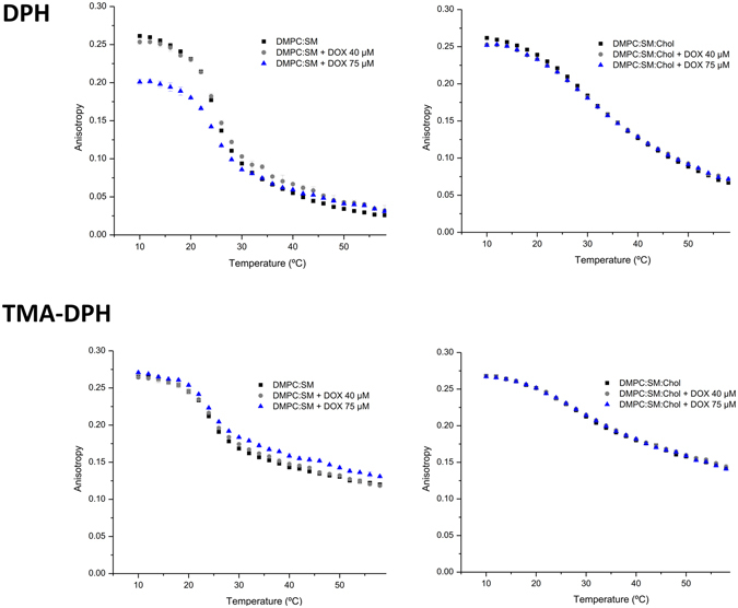
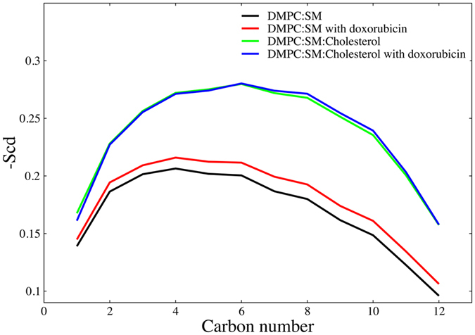
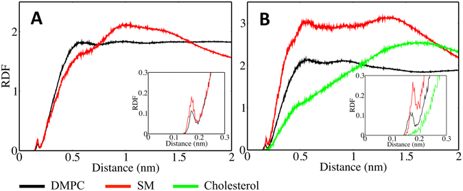
Despite doxorubicin being commonly used in chemotherapy there still remain significant holes in our knowledge regarding its delivery efficacy and an observed resistance mechanism that is postulated to involve the cell membrane. One possible mechanism is the efflux by protein P-gp, which is found predominantly in cholesterol enriched domains. Thereby, a hypothesis for the vulnerability of doxorubicin to efflux through P-gp is its enhanced affinity for the ordered cholesterol rich regions of the plasma membrane. Thus, we have studied doxorubicin's interaction with model membranes in a cholesterol rich, ordered environment and in liquid-disordered cholesterol poor environment. We have combined three separate experimental protocols: UV-Vis spectrophotometry, fluorescence quenching and steady-state anisotropy and computational molecular dynamics modeling. Our results show that the presence of cholesterol induces a change in membrane structure and doesn't impair doxorubicin's membrane partitioning, but reduces drug's influence on membrane fluidity without directly interacting with it. It is thus possible that the resistance mechanism that lowers the efficacy of doxorubicin, results from an increased density in membrane regions where the efflux proteins are present. This work represents a successful approach, combining experimental and computational studies of membrane based systems to unveil the behavior of drugs and candidate drug molecules.


Conflict of interest statement

The authors declare that they have no competing interests.

Figures

Figure 1
Figure 1
Molecular structure and pKa value of doxorubicin.
Figure 2
Figure 2
Third-derivative absorption spectra (A) of doxorubicin (40 μM) (red line, 0) alone, incubated in DMPC:SM model membrane at 37 °C with increasing lipid concentration (7 represents the maximum lipid concentration) and the model membrane without drug (black lines). (B) represents the best fitting curve to experimental third-derivative spectrophotometric data (DT vs. [L]) using a nonlinear regression method at a wavelength of 544 nm.
Figure 3
Figure 3
Stern–Volmer plots of the probe DPH in DMPC:SM [8:2] model at pH 7.4 and 37 °C with increasing doxorubicin concentrations: square symbols (◾) represent the Stern–Volmer plot obtained by steady-state fluorescence measurements (I0/I−1) and circle symbols (formula image
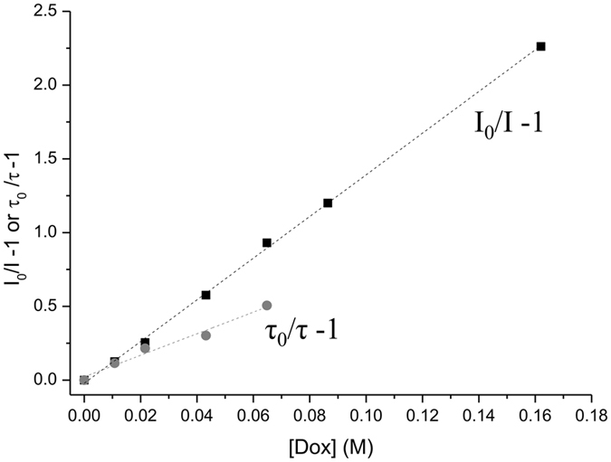
) represent the Stern–Volmer plot obtained by lifetime fluorescence measurements (τ0/τ−1).
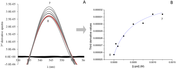
Figure 4
Figure 4
Steady-state anisotropy of DPH and TMA-DPH as a function of temperature in DMPC:SM [8:2] and DMPC:SM:Chol [7:1.5:1.5] membrane models. Results present the mean of at least three independent assays.
Figure 5
Figure 5
Steady-state anisotropy of DPH and TMA-DPH as a function of temperature in each mimetic model, in the absence (◾), and in the presence of doxorubicin 40 µM (formula image) and 75 µM (formula image). Results present the mean of at least three independent assays.
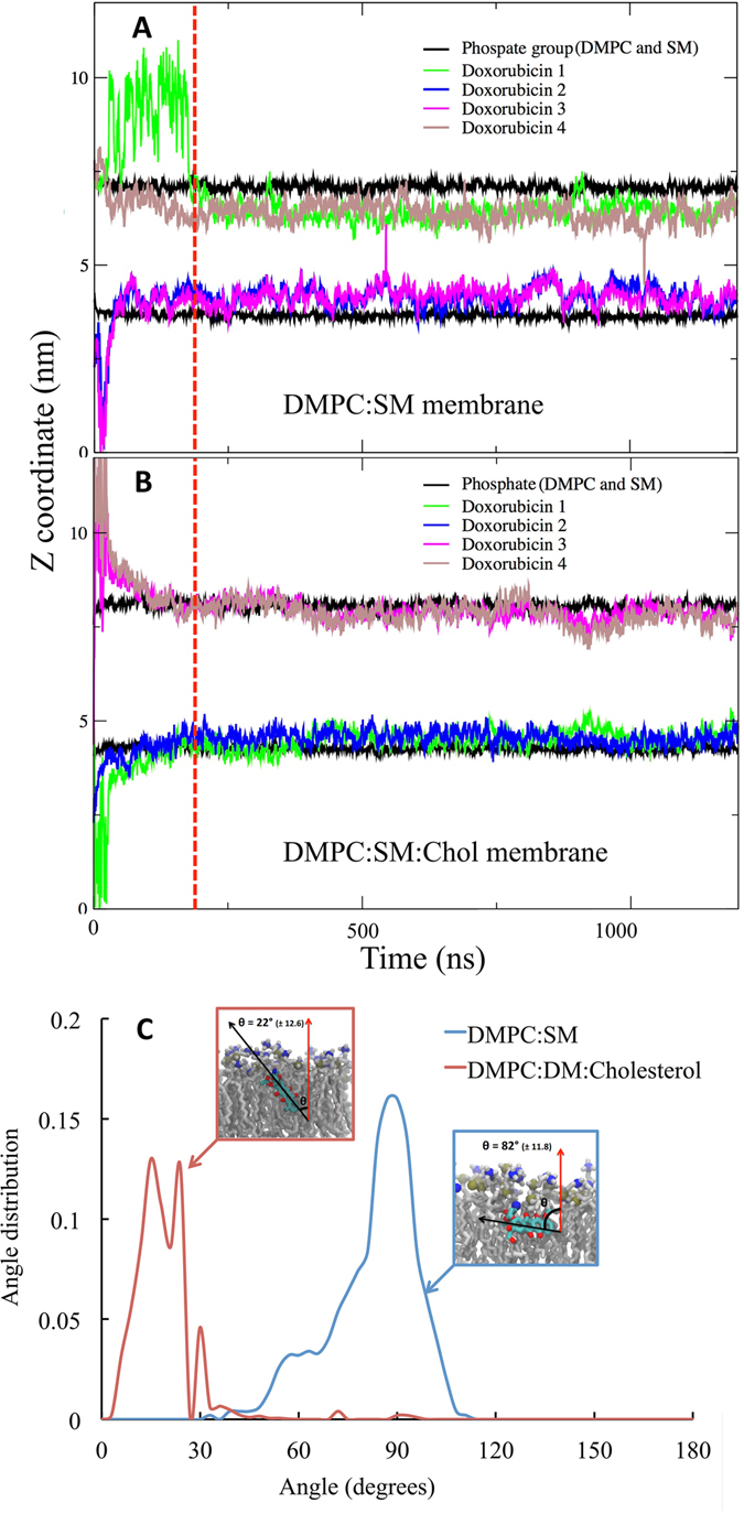
Figure 6
Figure 6
Partitioning of doxorubicin into the membrane bilayer models. The plot shows z-coordinate vs time for the center of mass of the phosphate group (of DMPC and SM) and doxorubicin molecules for (A) DMPC:SM [8:2] and (B) DMPC:SM:Chol [7:1.5:1.5] membranes. The red dotted line represents time point where systems were considered to be equilibrated, and for all analysis trajectory past this point (200 ns) was used. (C) The Angle distribution of doxorubicin in with membrane normal, in DMPC:SM (blue) and DMPC:SM:Chol (red) membranes. The error values represented are calculated as block errors.
Figure 7
Figure 7
Order parameter of the sn-2 chain of DMPC molecule. Order parameter shows that the increase in lipid order parameter in DMPC:SM membrane is more pronounced as compared to DMPC:SM:Chol membrane in presence of doxorubicin molecule.
Figure 8
Figure 8
Molecular interactions of doxorubicin with individual components of the membrane bilayers: Radial distribution function of the DMPC, SM headgroups and cholesterol OH group from doxorubicin. (A) DMPC:SM [8:2] with doxorubicin and (B) DMPC:SM:Chol [7:1.5:1.5] with doxorubicin.

References

    1. Minotti G, Menna P, Salvatorelli E, Cairo G, Gianni L. Anthracyclines: molecular advances and pharmacologic developments in antitumor activity and cardiotoxicity. Pharmacol. Rev. 2004;56:185–229. doi: 10.1124/pr.56.2.6. - DOI - PubMed
    1. Triton TR, Yee G. The anticancer agent adriamycin can be actively cytotoxic without entering cells. Science. 1982;217:248–250. doi: 10.1126/science.7089561. - DOI - PubMed
    1. Tritton, T. R. & Hickman, J. A. In Experimental and Clinical Progress in Cancer Chemotherapy24, 81–131 (Springer US, 1985).
    1. Escribá PV, et al. Membranes: a meeting point for lipids, proteins and therapies. J. Cell. Mol. Med. 2008;12:829–875. doi: 10.1111/j.1582-4934.2008.00281.x. - DOI - PMC - PubMed
    1. Escribá PV, et al. Role of lipid polymorphism in G protein-membrane interactions: nonlamellar-prone phospholipids and peripheral protein binding to membranes. Proceedings of the National Academy of Sciences. 1997;94:11375–11380. doi: 10.1073/pnas.94.21.11375. - DOI - PMC - PubMed

Publication types

MeSH terms

Substances