Case Reports
. 2017 Jul 4:82:364-370.
doi: 10.12659/PJR.901602. eCollection 2017.

Salvage Embolization and Histologic Analysis of Gastric Cancer with Hemorrhagic Shock Using Spherical Embolic Material

Affiliations
Case Reports

Salvage Embolization and Histologic Analysis of Gastric Cancer with Hemorrhagic Shock Using Spherical Embolic Material

Norifumi Kennoki et al. Pol J Radiol. .

Abstract

Background: In a recent study, it was reported that transcatheter arterial embolization with spherical embolic material for life-threatening hemorrhages in various cancer patients was safe and effective. Calibrated microspheres are able to access distal regions of the target arteries, which results in the disappearance of tumor staining. However, there are few reports on the pathological behavior of EmboSpheres in gastric cancer specimens. In this case, we succeeded in salvage embolization for advanced gastric cancer with hemorrhagic shock using spherical embolic material. To our knowledge, this is the first report of a pathological evaluation of spherical embolic microspheres in a gastric cancer specimen.

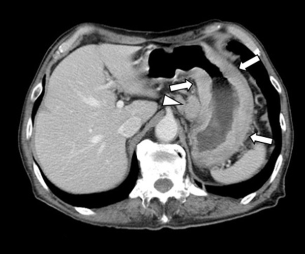
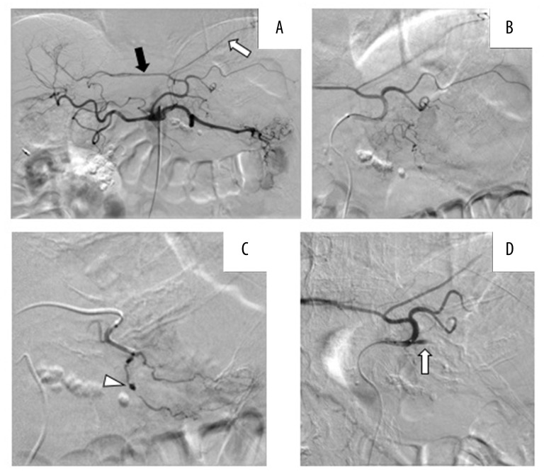
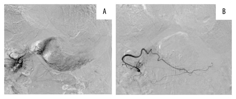
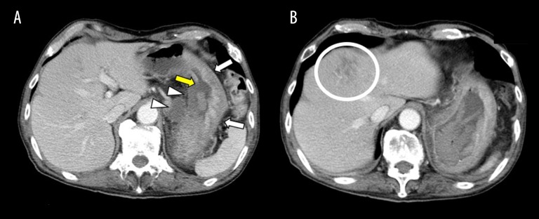
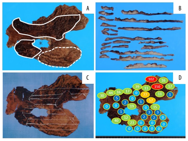
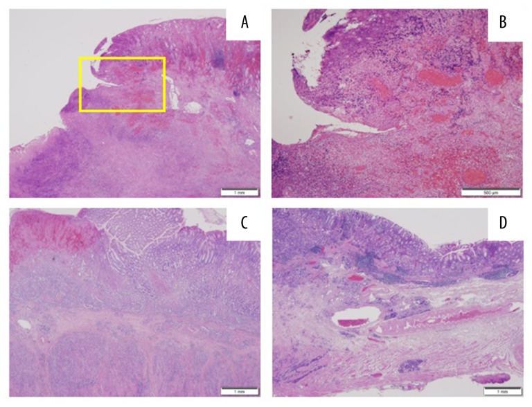
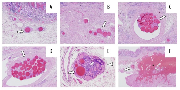
Case report: A 70-year-old man with scirrhous gastric cancer was admitted to our hospital for staging laparoscopy. Unfortunately, he had a sudden onset of hematemesis and melena leading to hemorrhagic shock due to bleeding from the gastric cancer. While undergoing a rapid blood transfusion, he underwent emergent embolization to achieve hemostasis. The left gastric and right gastroepiploic arteries were embolized with spherical embolic material, and the patient survived. Two days later, the patient was able to undergo gastrectomy. A large number of microspheres were observed in areas of hemorrhage. The range and median diameter of the minor axis were 177-1048 μm and 281 μm, respectively.

Conclusions: Transcatheter arterial embolization using spherical embolic material could become one of safe and effective options, especially when there is no extravasation or pseudoaneurysm but only tumor staining from the clinical and pathological point of view.

Keywords: Catheterization, Peripheral; Embolization, Therapeutic; Hemostasis; Microspheres; Stomach Neoplasms.


Figures

Figure 1
Figure 1
Initial contrast-enhanced computed tomography images. Thickening of the entire circumference of the gastric body wall (arrows) was displayed. A swollen lymph node was displayed in the lesser curvature (arrowhead).
Figure 2
Figure 2
Digital subtraction angiography (DSA) images of the celiac artery and left gastric artery. (A) Selective celiac artery angiography. The left hepatic artery (black arrow) and left subphrenic artery (white arrow) branched from the left gastric artery. (B) Selective left gastric artery angiography. (C) The pseudoaneurysm was located on a branch from the left gastric artery (arrowhead). (D) After the embolization using gelatin sponge and EmboSpheres the target branch was occluded completely.
Figure 3
Figure 3
DSA images of the right gastroepiploic artery. (A) Although extravasation and pseudoaneurysm were not displayed, the tumor staining was seen along the gastric wall in the greater curvature. (B) After embolization using EmboSpheres only, final angiography showed disappearance of tumor staining and the remaining main trunk of the right gastroepiploic artery (arrow).
Figure 4
Figure 4
Contrast-enhanced computed tomography images. (A) Two days after embolization neither the gastric body wall nor the swollen lymph node in the lesser curvature were enhanced (arrowheads). On the other hand, the gastric body wall in the greater curvature remained enhanced (arrows). An area of the gastric wall mucosa membrane in the lesser curvature was collapsed (yellow arrows). (B) A low-density area was located in segment 4 of the liver, indicating infarction (circle).
Figure 5
Figure 5
Gastric cancer specimen. (A) The area of the ulcer that adhered to and remained on the patient’s pancreas (asterisk), areas of hemorrhage (surrounded by solid lines) and a thick gastric wall (region surrounded by the dotted line) were macroscopically observed. (B) Cross-section after sectioning showed thick and white regions, which indicated cancer cell infiltration. (C) The spread of cancer cells is indicated with white lines. (D) The number of EmboSpheres detected in each region of the specimen.
Figure 6
Figure 6
Pathological findings on hematoxylin and eosin staining. (A) The lesion due to the ulcer near the mucosa formed a slope. (B) High-power field of the area within the square in (A). Hemorrhage, venous stasis and necrotic cancer cells were observed in the submucosal layer. (C) The posterior wall in the greater curvature – numerous and dense cancer cells were observed in the submucosa and between the muscles. (D) The anterior wall in the greater curvature - few cancer cells were observed compared with the posterior wall.
Figure 7
Figure 7
Pathological images showing embolic agents embolizing all arteries in the submucosa of the gastric wall. (A) An EmboSpehre. (B) 6 EmboSpheres. (C) 19 EmboSpheres. (D) 42 EmboSpheres. (E) EmboSpheres (arrows) and gelatin sponge (arrow head) embolizing one artery. (F) EmboSpheres (arrow), embolization and hemorrhage (asterisk).

References

    1. Lee HH, Park JM, Chun HJ, et al. Transcatheter arterial embolization for endoscopically unmanageable non-variceal upper gastrointestinal bleeding. Scand J Gastroenterol. 2015;50(7):809–15. - PubMed
    1. So JBY, Yam A, Cheah WK, et al. Risk factors related to operative mortality and morbidity in patients undergoing emergency gastrectomy. Br J Surg. 2000;87(12):1702–7. - PubMed
    1. Kim YI, Choi IJ. Endoscopic management of tumor bleeding from inoperable gastric cancer. Clin Endosc. 2015;48(2):121–27. - PMC - PubMed
    1. Loffroy R, Rao P, Ota S, et al. Embolization of acute nonvariceal upper gastrointestinal hemorrhage resistant to endoscopic treatment: Results and predictors of recurrent bleeding. Cardiovasc Intervent Radiol. 2010;33(6):1088–100. - PubMed
    1. Koh KH, Kim K, Kwon DH, et al. The successful endoscopic hemostasis factors in bleeding from advanced gastric cancer. Gastric Cancer. 2013;16(3):397–403. - PubMed

Publication types

LinkOut - more resources