. 2017 Apr-Jun;9(2):59-66.

Structure Modeling of Human Tyrosyl-DNA Phosphodiesterase 1 and Screening for Its Inhibitors

Affiliations

Structure Modeling of Human Tyrosyl-DNA Phosphodiesterase 1 and Screening for Its Inhibitors

I V Gushchina et al. Acta Naturae. 2017 Apr-Jun.

Abstract

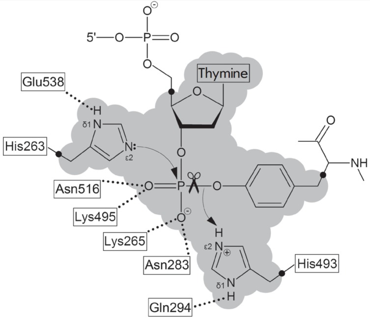
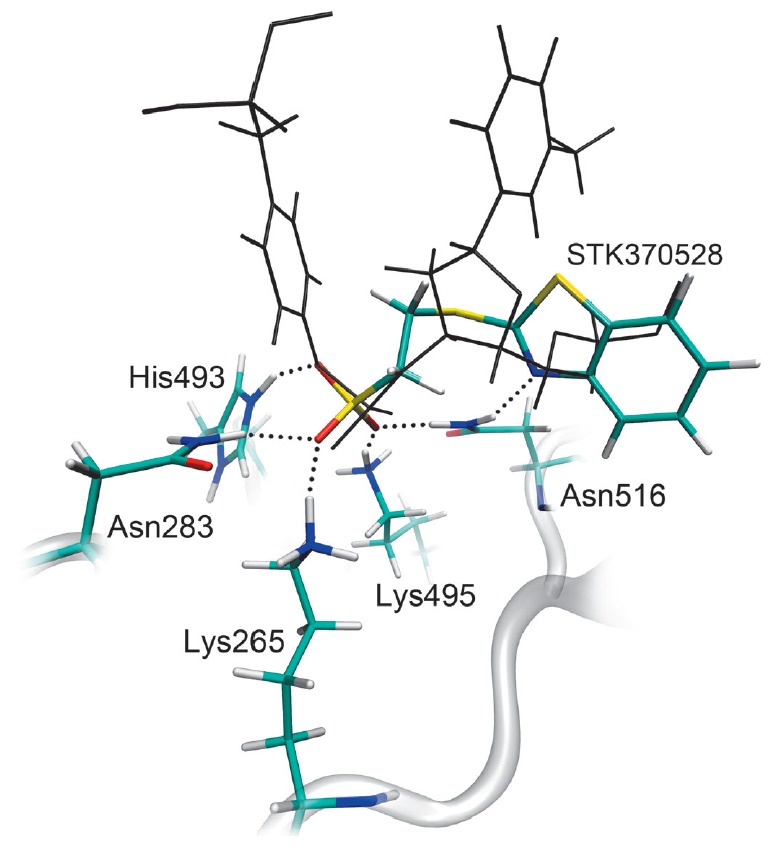
The DNA repair enzyme tyrosyl-DNA phosphodiesterase 1 (Tdp1) represents a potential molecular target for anticancer therapy. A human Tdp1 model has been constructed using the methods of quantum and molecular mechanics, taking into account the ionization states of the amino acid residues in the active site and their interactions with the substrate and competitive inhibitors. The oligonucleotide- and phosphotyrosine-binding cavities important for the inhibitor design have been identified in the enzyme's active site. The developed molecular model allowed us to uncover new Tdp1 inhibitors whose sulfo group is capable of occupying the position of the 3'-phosphate group of the substrate and forming hydrogen bonds with Lys265, Lys495, and other amino acid residues in the phosphotyrosine binding site.

Keywords: docking; inhibitor; molecular modeling; tyrosyl-DNA phosphodiesterase 1.


Figures

Fig. 1
Fig. 1
The structure of the Tdp1 active site. The Lys265, Asn283, Lys495, and Asn516 residues are involved in the binding of the substrate’s phosphate group. In the reaction mechanism, nucleophilic attack by His263 residue occurs and a proton is transferred from His493 to the leaving group. The shaded area corresponds to the QM region defined in the performed modeling of the enzyme-substrate complex.
Fig. 2
Fig. 2
The substrate-binding groove in the human Tdp1 model. (A) The interaction of the substrate molecule with the oligonucleotide and phosphotyrosine binding sites. The oligonucleotide is shown in red, phosphotyrosine is shown in green, and the rest of the peptide is shown in yellow. Cavities are labeled with points corresponding to the centers of alpha spheres. (B) The localization of alpha spheres in the oligonucleotide and phosphotyrosine binding sites.
Fig. 3
Fig. 3
Interactions of inhibitors with the substrate-binding groove in the human Tdp1 model. (A) The binding of diazaadamantane derivatives. (B) The binding of the sulfo-substituted derivatives STK370528 and STK376552. The oligonucleotide and phosphotyrosine cavities are labeled with red and green alpha spheres, respectively.
Fig. 4
Fig. 4
The dependence of the Tdp1-catalyzed reaction rate on the concentration of the inhibitor STK370528.
Fig. 5
Fig. 5
The position of the inhibitor, STK370528, in the active site of the molecular model of human Tdp1. Dotted lines indicate hydrogen bonds important for the stabilization of the sulfo group position. The gray color denotes the substrate coordinates in the model of the enzyme-substrate complex.

References

    1. Champoux J.J.. Annu. Rev. Biochem. 2001;70:369–413. - PubMed
    1. Wang J.C.. Nat. Rev. Mol. Cell Biol. 2002;3:430–440. - PubMed
    1. Pommier Y.. Nat. Rev. Cancer. 2006;6:789–802. - PubMed
    1. Lebedeva N., Rechkunova N., Boiteux S., Lavrik O.. IUBMB Life. 2008;60:130–134. - PubMed
    1. Pommier Y., Redon C., Rao V.A., Seiler J.A., Sordet O., Takemura H., Antony S., Meng L., Liao Z., Kohlhagen G.. Mutat. Res. 2003;532:173–203. - PubMed

LinkOut - more resources