. 2018 Jan;223(1):165-182.
doi: 10.1007/s00429-017-1476-1. Epub 2017 Jul 24.

Shifted dynamic interactions between subcortical nuclei and inferior frontal gyri during response preparation in persistent developmental stuttering

Affiliations

Shifted dynamic interactions between subcortical nuclei and inferior frontal gyri during response preparation in persistent developmental stuttering

F Luise Metzger et al. Brain Struct Funct. 2018 Jan.

Abstract

Persistent developmental stuttering is associated with basal ganglia dysfunction or dopamine dysregulation. Here, we studied whole-brain functional connectivity to test how basal ganglia structures coordinate and reorganize sensorimotor brain networks in stuttering. To this end, adults who stutter and fluent speakers (control participants) performed a response anticipation paradigm in the MRI scanner. The preparation of a manual Go/No-Go response reliably produced activity in the basal ganglia and thalamus and particularly in the substantia nigra. Strikingly, in adults who stutter, substantia nigra activity correlated positively with stuttering severity. Furthermore, functional connectivity analyses yielded altered task-related network formations in adults who stutter compared to fluent speakers. Specifically, in adults who stutter, the globus pallidus and the thalamus showed increased network synchronization with the inferior frontal gyrus. This implies dynamic shifts in the response preparation-related network organization through the basal ganglia in the context of a non-speech motor task in stuttering. Here we discuss current findings in the traditional framework of how D1 and D2 receptor activity shapes focused movement selection, thereby suggesting a disproportional involvement of the direct and the indirect pathway in stuttering.

Keywords: Basal ganglia; Disinhibition; Inferior frontal gyrus; Persistent developmental stuttering; Response anticipation; Substantia nigra.


Figures
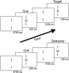
Fig. 1
Fig. 1
Schematic illustration of the continuous performance task. The three letters “H”, “O”, or “X” served as stimuli. Participants were asked to respond with a right-hand button press if the cue letter (O) was followed by the target letter (X). If the cue letter (O) was followed by any other letter (H, O), participants had to suppress the prepared motor response. The design was adopted from Lütcke et al. (2008)
Fig. 2
Fig. 2
Definition of the seed region in the substantia nigra (SN). To extract physiological time courses of the SN, bilateral masks were manually drawn in the mean EPIs of every run of each participant. Four examples are displayed for control participants and adults who stutter (AWS), respectively. Right columns display the contrast of the mean EPI, left rows display SN masks overlaid in red. The axial brain slice in the right column of the figure illustrates the overlap of substantia nigra seed regions in the current data set across all 27 participants, aligned with the 2-mm MNI standard brain
Fig. 3
Fig. 3
Results of the CPT. The upper panel histograms display all reaction times, separated for groups illustrating a broad overlap. For reaction times, box plots were calculated across median reaction times, separated for group and run. For accuracy, box plots were calculated across the pooled data. Both groups performed comparably well and only showed a few false alarms and omissions, resulting in high d′ scores. Whiskers display the 10th and 90th percentile. Maps in the middle panel show brain activations during response anticipation. The contrast was obtained by the comparison of activity following cues and non-cues across all participants. Anticipatory processes activated a large-scale cortical network including the frontal pole (FP), superior frontal gyrus (SFG), SMA, insula, inferior frontal gyrus pars opercularis (BA44), premotor cortex (pMC), motor cortex (M1), and somatosensory cortex (S1) together with the paracingulate gyrus (PCG) and anterior cingulate cortex (ACC). The broad activation of the basal ganglia and brainstem involving the caudate nucleus (CN), putamen, globus pallidus (GP), thalamus, red nucleus (RN), subthalamic nucleus (STN) and also the substantia nigra (SN) is most prominent. Z statistic images were thresholded using clusters determined by Z > 3.1 and a (corrected) cluster significance threshold of p = 0.05 (Worsley 2001)
Fig. 4
Fig. 4
Region of interest analysis. The axial sections display the locations of the ROI masks of the substantia nigra (SN), external segment of the globus pallidus (GPe), and medial dorsal nucleus of the thalamus (MD). Box plots illustrate the BOLD activity during response anticipation separated for adults who stutter and control participants. Whiskers display the 10th and 90th percentile. Scatter plots show the relationship between BOLD activity and stuttering severity. Stuttering severity as indicated by the SSI-4 score was correlated positively with the task-related activation of the right SN. Marker sizes in the scatter plots indicate percent stuttered syllables with largest diameters for highest frequencies
Fig. 5
Fig. 5
PPI results across adults who stutter and control participants during response anticipation seeding in the medial dorsal nucleus of the left thalamus (MD) and the external segment of the globus pallidus (GPe). Z statistic images were thresholded using clusters determined by Z > 2.3 and a corrected cluster significance threshold of p = 0.05 (Worsley 2001)
Fig. 6
Fig. 6
PPIs resulted in group differences when seeding in the medial dorsal nucleus of the left thalamus (MD) and when seeding in the external segment of the globus pallidus (GPe). Z statistic images were thresholded using clusters determined by Z > 2.3 and a corrected cluster significance threshold of p = 0.05 (Worsley 2001)
Fig. 7
Fig. 7
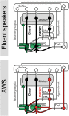
Cortico–basal ganglia–thalamo–cortical loops. The diagram displays a simplified model of the direct, indirect, and hyperdirect pathway. White indicates glutamatergic (excitatory) connections, black indicates GABAergic (inhibitory) connections, and green indicates dopaminergic (modulatory) connections. Adults who stutter exhibited altered network dynamics between cortex and the external segment of the globus pallidus (GPe). For this reason, the GPe and its downstream structures are marked in red. Remarkably, the GPe is a principal structure of the indirect pathway

Similar articles

Cited by

References

    1. Alexander GE, Crutcher MD. Functional architecture of basal ganglia circuits: neural substrates of parallel processing. Trends Neurosci. 1990;13:266–271. doi: 10.1016/0166-2236(90)90107-L. - DOI - PubMed
    1. Allert N, Kelm D, Blahak C, et al. Stuttering induced by thalamic deep brain stimulation for dystonia. J Neural Transm. 2010;117:617–620. doi: 10.1007/s00702-010-0380-0. - DOI - PubMed
    1. Alm PA. Stuttering and the basal ganglia circuits: a critical review of possible relations. J Commun Disord. 2004;37:325–369. doi: 10.1016/j.jcomdis.2004.03.001. - DOI - PubMed
    1. Anderson J, Hughes J, Rothi L, et al. Developmental stuttering and Parkinson’s disease: the effects of levodopa treatment. J Neurol Neurosurg Psychiatry. 1999;66:776–778. doi: 10.1136/jnnp.66.6.776. - DOI - PMC - PubMed
    1. Bandettini PA, Wong EC, Jesmanowicz A, et al. Spin-echo and gradient-echo EPI of human brain activation using BOLD contrast: a comparative study at 1.5 T. NMR Biomed. 1994;7:12–20. doi: 10.1002/nbm.1940070104. - DOI - PubMed

MeSH terms