Review
. 2015 Sep;1(1):76-91.
doi: 10.1016/j.trecan.2015.07.009. Epub 2015 Sep 28.

Surviving at a Distance: Organ-Specific Metastasis

Affiliations
Review

Surviving at a Distance: Organ-Specific Metastasis

Anna C Obenauf et al. Trends Cancer. 2015 Sep.

Abstract

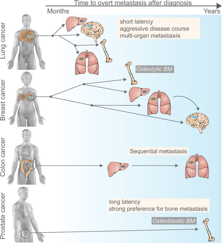
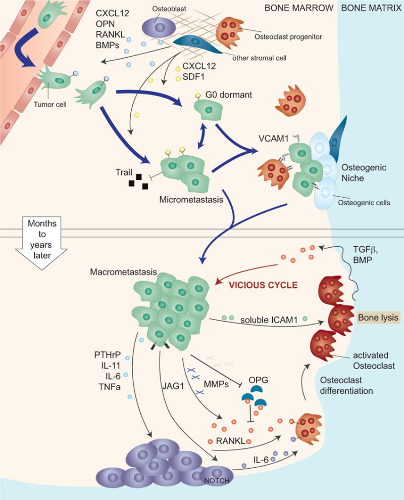
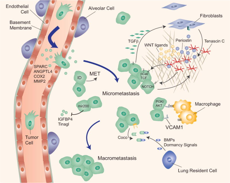
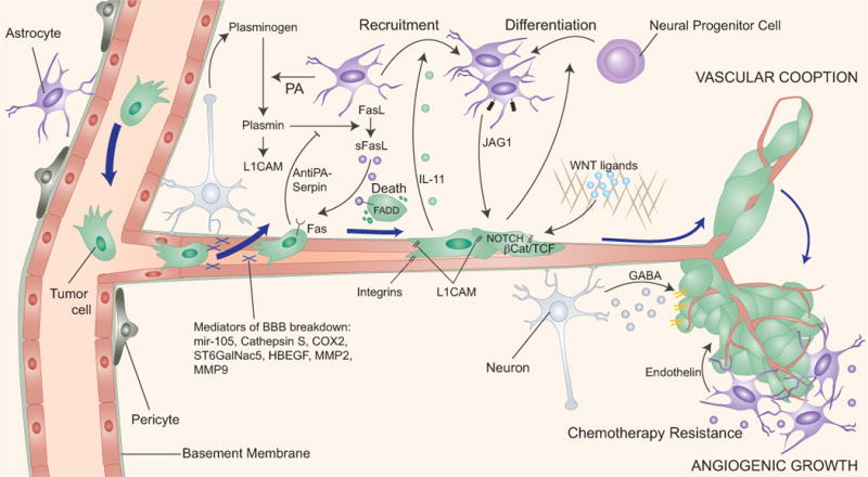
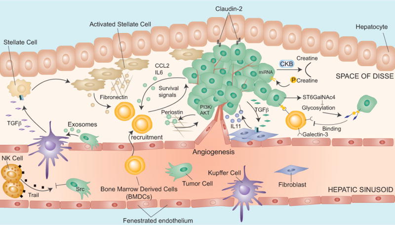
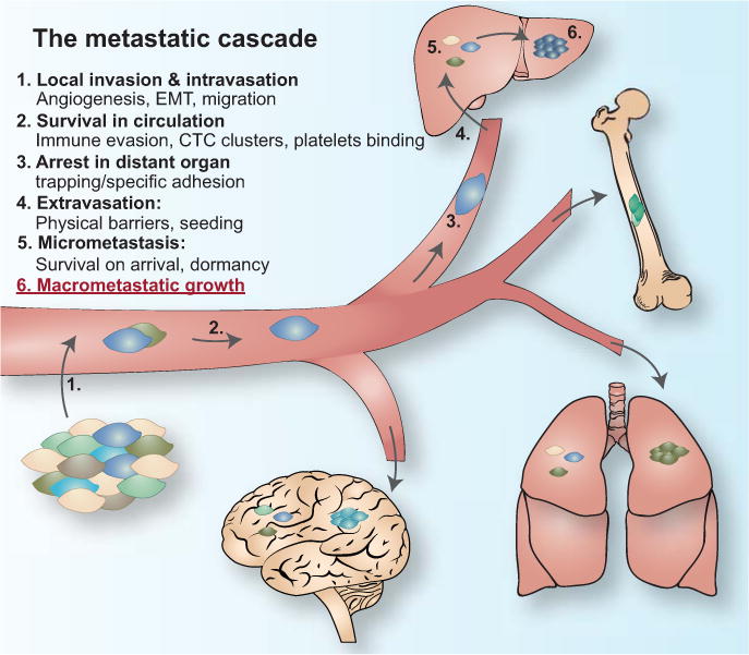
The clinical manifestation of metastasis in a vital organ is the final stage of cancer progression and the main culprit of cancer-related mortality. Once established, metastasis is devastating, but only a small proportion of the cancer cells that leave a tumor succeed at infiltrating, surviving, and ultimately overtaking a distant organ. The bottlenecks that challenge cancer cells in newly invaded microenvironments are organ-specific and consequently demand distinct mechanisms for metastatic colonization. We review the metastatic traits that allow cancer cells to colonize distinct organ sites.

Keywords: Cancer; metastasis; metastatic colonization; organ-specific metastasis.


Figures

Figure 1, Key Figure
Figure 1, Key Figure. Patterns of metastatic spread of solid tumors
Different cancer types exhibit remarkable variability in their metastatic course, reflected in the length of the latency period (months to years), the affected organs (most commonly the liver, lung, bone, and brain) and the type of metastasis (e.g. osteolytic or osteoblastic bone metastasis). Latency period (denoted by the arrow on top of the figure, left: months, right: years after diagnosis): Lung cancer metastasis typically occurs within months after initial diagnosis, whereas prostate cancer and certain subtypes of breast cancer can produce distant relapse up to decades after initial diagnosis. Lung cancer is the main contributor to brain metastasis, a late occurrence in breast cancer. Organ pattern (most frequently affected organ is located on the top of each cancer type): Lung and breast cancers metastasize to different organs (with a different propensity), whereas colon cancer most frequently metastasizes to liver, and from established liver metastasis secondarily to lung. Prostate cancer typically, though not exclusively metastasizes to bone. Different cancer types also vary in the type of metastatic lesions they induce, well illustrated by the development of osteolytic bone metastasis in breast and lung cancer and osteoblastic bone metastasis in prostate cancer.
Figure 2
Figure 2. Osteolytic metastatic colonization of the bone
The capillaries in the bone, called sinusoids, are lined with fenestrated endothelia that facilitate the traffic of hematopoietic cells. Thus, the bone marrow sinusoids are likely permissive to cancer cell passage. Upper panel: Upon infiltrating the bone marrow cancer cells are exposed to a variety of growth and death promoting signals, which are thought to force cancer cells into a latent state until they acquire the necessary traits for overt metastasis. In this state cancer cells benefit from secreted survival signals (CXCL12, SDF1) from bone resident cells and by direct interaction with osteogenic cells and pre-osteoclasts. Lower panel: A critical step in the formation of overt osteolytic bone metastasis is the activation of osteoclasts. This process is locally facilitated by cancer cell-derived mediators including PTHrP, IL-11 and others that stimulate the secretion of RANKL by osteoblasts. Cleavage and release of membrane bound RANKL, or inactivation of the antagonist osteoprotegerin (OPG) can also contribute to increasing RANKL activity. Alternatively, cancer cells trigger the secretion of IL-6 by osteoblasts, which in turn induces osteoclast differentiation. Activated osteoclasts execute bone resorption, which releases TGF-β and other growth factors that are embedded in the mineralized bone matrix. TGF-β then further stimulates the expression of osteolytic factors in the cancer cells, resulting in a vicious cycle of bone metastasis.
Figure 3
Figure 3. Metastatic colonization of the lung
The lung capillaries are lined with a basement membrane and mediators of cancer cell extravasation in the lung have been identified (e.g. SPARC, ANGPTL4, COX2, MMP2). The expression of ID proteins in breast cancer cells induces mesenchymal-epithelial transition (MET) at the metastatic site and supports in breast cancer cells in bypassing senescence. The cancer cell-and myofibroblast-derived extracellular matrix proteins periostin (POSTN) and tenascin C (TNC) stimulate cancer cell survival by enhancing Wnt access to their receptors (periostin) or by amplifying Wnt and Notch signaling (TNC). Cancer cells express high levels of vascular cell adhesion molecule 1 (VCAM1), which tethers macrophages to cancer cells and triggers activation of the cell survival AKT pathway in cancer cells. Cancer cells overcome dormancy signals (BMPs) from lung resident cells by overexpressing Coco, which increases formation of macrometastasis.
Figure 4
Figure 4. Metastatic colonization of the brain
To establish parenchymal brain metastasis cancer cells have to cross the vascular walls that constitute the blood brain barrier (BBB), which consists of tightly connected endothelial cells lined with a basement membrane and contacting astrocyte and pericytes. Several classes of mediators of cancer cell passage through the BBB have been identified (mir-105, Cathepsin S, COX2, ST6GalNac5, HBEGF, MMP2, MMP9). Cancer cells express high levels of anti-PA serpins which prevent the release of cytotoxic soluble Fas-ligand (sFasl) from reactive astrocytes and the inactivation of the L1CAM adhesion molecule that mediates vascular cooption by the cancer cells. Once cancer cells evade astrocyte-mediated killing they can take advantage of astrocyte-derived survival and chemo-protective functions of largely unknown nature. Cancer cells may stimulate the accumulation of astrocytes in metastatic lesions. Cancer cells may also utilize neuron-secreted GABA as support for metastasis.
Figure 5
Figure 5. Metastatic colonization of the liver
The extravasation into the liver is facilitated by the hepatic vascular endothelium, which is fenestrated and lacks an organized basement membrane. High Src signaling protects cancer cells from TRAIL-mediated apoptosis. Cancer cells release MIF containing exosomes that trigger TGF-β production for the activation of stellate cells, leading to the recruitment of bone marrow derived cells (BMDCs). BMDCs can also be attracted by secretion of CCL2 and IL6. Other survival signals may be provided by galectin-3. In the liver, colon cancer cells secrete periostin, which induces PI3K/AKT signaling. Cancer cells also interact with hepatocytes via claudin-2, stimulating overt metastasis. The secretion of creatine kinase B (CKB) by cancer cells contributes to metastatic outgrowth by generating phosphocreatine as a metabolite to regenerate ATP in cancer cells.
Figure I for Text Box 1
The metastatic cascade

References

    1. Nguyen DX, Bos PD, Massague J. Metastasis: from dissemination to organ-specific colonization. Nat Rev Cancer. 2009;9(4):274–84. - PubMed
    1. Valastyan S, Weinberg RA. Tumor metastasis: molecular insights and evolving paradigms. Cell. 2011;147(2):275–92. - PMC - PubMed
    1. Steeg PS. Tumor metastasis: mechanistic insights and clinical challenges. Nat Med. 2006;12(8):895–904. - PubMed
    1. Disibio G, French SW. Metastatic patterns of cancers: results from a large autopsy study. Arch Pathol Lab Med. 2008;132(6):931–9. - PubMed
    1. Budczies J, et al. The landscape of metastatic progression patterns across major human cancers. Oncotarget. 2015;6(1):570–83. - PMC - PubMed