. 2017 Sep;24(9):752-757.
doi: 10.1038/nsmb.3439. Epub 2017 Jul 24.

An antimicrobial peptide that inhibits translation by trapping release factors on the ribosome

Affiliations

An antimicrobial peptide that inhibits translation by trapping release factors on the ribosome

Tanja Florin et al. Nat Struct Mol Biol. 2017 Sep.

Abstract

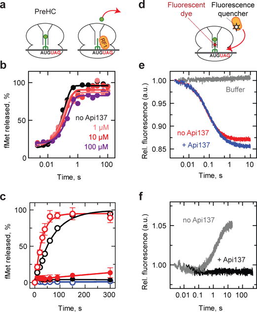
Many antibiotics stop bacterial growth by inhibiting different steps of protein synthesis. However, no specific inhibitors of translation termination are known. Proline-rich antimicrobial peptides, a component of the antibacterial defense system of multicellular organisms, interfere with bacterial growth by inhibiting translation. Here we show that Api137, a derivative of the insect-produced antimicrobial peptide apidaecin, arrests terminating ribosomes using a unique mechanism of action. Api137 binds to the Escherichia coli ribosome and traps release factor (RF) RF1 or RF2 subsequent to the release of the nascent polypeptide chain. A high-resolution cryo-EM structure of the ribosome complexed with RF1 and Api137 reveals the molecular interactions that lead to RF trapping. Api137-mediated depletion of the cellular pool of free release factors causes the majority of ribosomes to stall at stop codons before polypeptide release, thereby resulting in a global shutdown of translation termination.


Conflict of interest statement

Competing financial interests:

The authors declare no competing financial interests.

Figures

Figure 1
Figure 1. Api137 stalls ribosomes at the termination step of translation
a, Amino acid sequences of PrAMPs Api137 and Onc112. gu = N,N,N′,N′-tetramethylguanidino, O = L-ornithine, r = D-arginine. b, c, In vitro toeprinting analysis comparing the Onc112- or Api137- mediated translation arrest on model mRNA templates derived from the yrbA (b) or ermCL (c) genes. Positions of the toeprint bands (indicated on the gene sequence) are 16–17 nt downstream from the first nucleotide of the P-site codon. The P- and A- sites codons of the stalled ribosomes are in brackets. Toeprints in (c) were produced by wild-type ribosomes (wt) or by ribosomes with mutations in specific rRNA nucleotides (Supplementary Fig. 2a). Toeprint bands in (b) and (c) generated by Onc112-arrested ribosomes at the initiation codon are indicated with grey arrowheads; those from ribosomes arrested by Api137 at termination are marked with white arrowheads. The similar intensity of the PrAMP-independent toeprint bands marked with a white arrowhead with dotted outline in (c) shows that wt and mutant ribosomes translate with comparable efficiencies. Sequencing reactions are marked. The gels are representatives of (b) more than five and (c) two independent biological replicates.
Figure. 2
Figure. 2. Api137 allows peptide hydrolysis but inhibits turnover of RF1 and RF2
a, Schematics of the peptidyl-tRNA hydrolysis experiments. PreHC carrying f[3H]Met-tRNAfMet is reacted with RF1 (shown) or RF2 and the release of f[35S]Met is measured. b, Time courses of peptide hydrolysis in PreHC in the presence of excess of RF1 without (black) or with the indicated concentrations of Api137 (colored traces). c, Time courses of peptide hydrolysis in PreHC by RF1 (black circles) and RF2 (red circles) under turnover conditions in the absence (open circles) or presence of 1 μM Api137. RF3-GTP was present in all reactions. Control experiments (blue circles) lacked RF1 and RF2 in the absence (open circles) or presence (closed circles) of Api137. 100% corresponds to 10 cycles of RFs binding, catalysis and dissociation. Error bars represent the range of two independent replicates. d, Schematics of the RF1 binding experiments. PreHC carries fluorescein-labeled fMet-tRNA (PreHCFlu) and RF1 carries fluorescence quencher dye (RF1Qsy). e, Time courses of binding of RF1Qsy to PreHCFlu in the absence (red) or presence (blue) or Api137. Grey trace: no RF1. The traces represent the average of five to seven technical replicates. f, Time course of RF1 dissociation. RF1Qsy was incubated with PreHCFlu to generate PostHCFlu and then mixed with a 10-fold excess of unlabeled RF1 and RF3·GTP in the absence (grey) or in the presence (black) of Api137. The traces represent the average of up to seven technical replicates. See Methods for details.
Figure. 3
Figure. 3. Binding of Api137 to the terminating ribosome and its interactions with the exit tunnel
a, Transverse section of the 50S subunit (grey) showing the binding site of Api137 (salmon) on the 70S ribosome (30S subunit in yellow) within the polypeptide exit tunnel relative to RF1 (orange) and the P-site tRNA (green). b, Cryo-EM density (mesh) and molecular model (salmon) for residues 5–18 of Api137. c, Placement of Api137 in the exit tunnel relative to RF1, P-site tRNA and ribosomal proteins uL4 and uL22. Boxed regions are zoomed in the panels d-f, showing interactions of Api137 with components of the exit tunnel, including (d, e) nucleotides of the 23S rRNA (grey) and (f) ribosomal protein uL4. In (e) sphere representation is used to approximate van der Waals interactions and in (f) a hydrogen bond is indicated with a dashed yellow line.
Figure 4
Figure 4. Inhibitory action of Api137 is mediated by its interactions with RF1 and P-site tRNA
a, Position of Api137 (salmon) relative to RF1 (orange) and P-site tRNA (green). The boxed regions are enlarged in panels (b,c). b, Interactions of Api137 with RF1. Arg17 of Api137 is coordinated by bonding with 23S rRNA nucleotides C2452, G2505 and U2506 (grey) to form direct contacts with Gln235 of the GGQ motif of RF1 (orange). c, The C-terminal hydroxyl of Leu18 of Api137 interacts with the ribose of A76 of deacylated P-site tRNA (green). d, Dimethylsulfate (DMS) probing of Api137 interaction with PostHC 23S rRNA in the absence or presence of RF1. This gel is a representative of two independent experiments.
Figure. 5
Figure. 5. Api137 induces accumulation of peptidyl-tRNA and stop codon read-through
a, Gel electrophoresis analysis of the [35S]-labeled products of the in vitro translation of the tnaC gene with its original UGA stop codon (lanes 1–6) or with the UAG stop codon (lanes 7–9), in the absence or presence of Api137. Where indicated, reaction products were treated with RNase. Lane 1, control reaction without mRNA. Lane 2 (labeled as M, marker) shows the band of RNase-sensitive TnaC-tRNA accumulated at high concentration of tryptophan. Lanes 5 and 8 show Api137-induced accumulation of TnaC-tRNA at low concentration of tryptophan. The bands of TnaC-tRNA and of the released TnaC peptide are shown with filled and open arrowheads, respectively. This gel is representative of five independent biological replicates. b, Excess of RF1 rescues Api137-induced accumulation of peptidyl-tRNA. Cell-free translation with low tryptophan was carried out in the standard conditions (lanes 1 and 3) or in the presence of a 5-fold molar excess of RF1 over the ribosomes (lane 2). This gel is representative of two independent biological replicates. c, In vivo expression of the chromosomal mutant lacZ with a premature stop codon mediated by the stop codon read-through stimulated by the miscoding antibiotic streptomycin (Str) or by Api137. The droplet of read-through inducing agents was placed at the indicated points on the lawn of the E. coli cells grown on an LB/agar plate supplemented with ampicillin, IPTG and X-Gal. This plate represents one of three independent experiments. d, e, The dual mode of Api137 action. d, Api137 binds to the ribosome after RF1 (or RF2) catalyzes the release of the complete protein and traps RF1 (or RF2) thereby preventing its turnover. e, Trapping of RF1 (or RF2) depletes their available pool causing stalling of most of the ribosomes at the stop codons unable to release the nascent proteins.

Similar articles

Cited by

References

    1. Korostelev AA. Structural aspects of translation termination on the ribosome. RNA. 2011;17:1409–1421. - PMC - PubMed
    1. Bremer H, Dennis P. In: Escherichia coli and Salmonella: Cellular and Molecular Biology. Neidhardt FC, et al., editors. Vol. 2. ASM Press; 1996. pp. 1553–1569. Ch. 97.
    1. Schmidt A, et al. The quantitative and condition-dependent Escherichia coli proteome. Nat Biotech. 2016;34:104–110. - PMC - PubMed
    1. Koutmou KS, McDonald ME, Brunelle JL, Green R. RF3:GTP promotes rapid dissociation of the class 1 termination factor. RNA. 2014;20:609–620. - PMC - PubMed
    1. Shi X, Joseph S. Mechanism of translation termination: RF1 dissociation follows dissociation of RF3 from the ribosome. Biochemistry. 2016;55:6344–6354. - PubMed

MeSH terms

LinkOut - more resources