. 2017 Aug;98(8):2030-2042.
doi: 10.1099/jgv.0.000859. Epub 2017 Jul 26.

Molecular cloning of porcine Siglec-3, Siglec-5 and Siglec-10, and identification of Siglec-10 as an alternative receptor for porcine reproductive and respiratory syndrome virus (PRRSV)

Affiliations

Molecular cloning of porcine Siglec-3, Siglec-5 and Siglec-10, and identification of Siglec-10 as an alternative receptor for porcine reproductive and respiratory syndrome virus (PRRSV)

Jiexiong Xie et al. J Gen Virol. 2017 Aug.

Abstract

In recent years, several entry mediators have been characterized for porcine reproductive and respiratory syndrome virus (PRRSV). Porcine sialoadhesin [pSn, also known as sialic acid-binding immunoglobulin-type lectin (Siglec-1)] and porcine CD163 (pCD163) have been identified as the most important host entry mediators that can fully coordinate PRRSV infection into macrophages. However, recent isolates have not only shown a tropism for sialoadhesin-positive cells, but also for sialoadhesin-negative cells. This observation might be partly explained by the existence of additional receptors that can support PRRSV binding and entry. In the search for new receptors, recently identified porcine Siglecs (Siglec-3, Siglec-5 and Siglec-10), members of the same family as sialoadhesin, were cloned and characterized. Only Siglec-10 was able to significantly improve PRRSV infection and production in a CD163-transfected cell line. Compared with sialoadhesin, Siglec-10 performed equally effectively as a receptor for PRRSV type 2 strain MN-184, but it was less capable of supporting infection with PRRSV type 1 strain LV (Lelystad virus). Siglec-10 was demonstrated to be involved in the endocytosis of PRRSV, confirming the important role of Siglec-10 in the entry process of PRRSV. In conclusion, it can be stated that PRRSV may use several Siglecs to enter macrophages, which may explain the strain differences in the pathogenesis.


Figures

Fig. 1.
Fig. 1.
Amino acid sequence and structure analysis of Siglec-1, Siglec-3, Siglec-5 and Siglec-10. (a) Spatial structure of Siglec-1,3, Siglec-5 and Siglec-10. Protein structure was predicted by I-TASSER (http://zhanglab.ccmb.med.umich.edu/I-TASSER/) and analysed by PyMOL (version 6.6). The yellow portion represents the signal peptide; the white portion represents the V-type Ig domain; and the red dots are the predicted sialic acid-binding site. Different numbers of C-set domains are shown just under the V-type Ig domain. The surface of the predicted sialic acid-binding sites was expanded and the numbers represent the amino acid position of the predicted sialic acid-binding sites. (b) Amino acid sequence homology analysis. The homology of the full-length Siglecs obtained in this study was compared with the sequences that have been reported using MegAlign (DNAstar). Highlighted numbers represent the similarity between the Siglecs obtained in this study with the reported ones. (c) Amino acid comparison of V-type Ig domain. The V-type Ig amino acid sequences of Siglec-1, -3, -5 and -10 were compared using CLC sequence viewer (version 6.8.1). The predicted sialic acid-binding site is indicated with an asterisk.
Fig. 2.
Fig. 2.
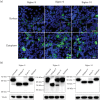
Expression analysis of Siglec-3, Siglec-5 and Siglec-10 using IFA and Western blotting. (a) PK-15 cells were transfected with Siglec-3-, Siglec-5- and Siglec-10-encoding vector. Twenty-four hours post-transfection, the cells were fixed with 4 % PF and permeabilized with Triton X-100 for cytoplasmic staining, or not permeabilized for surface staining. IFA was performed using V5-specific antibody (Siglec; green) and Hoechst 33 342 (nuclei; blue). Scale bar: 25 µm (b) Western blot identification. HEK-293T cells were transfected with Siglec-3, Siglec-5 and Siglec-10. Twenty-four hours post-transfection, the cells were collected and lysed with lysis buffer. Afterwards, cellular lysates were treated or mock-treated with sialidase, EndoH or PNGase F and analysed by SDS-PAGE and Western blot. Primary antibody V5-specific mAb (GenScript; A00641) and secondary peroxidase-labelled goat anti-mouse IgG antibodies (Dako) were used for immunoblotting. For the detection of tubulin, an HRP-conjugated anti-alpha tubulin monoclonal antibody (Abcam; ab40742) was used.
Fig. 3.
Fig. 3.
Analysis of the sialic acid-binding capacity of Siglec-1, Siglec-3, Siglec-5 and Siglec-10 by red blood cell binding assay. PK-15 cells that had been transiently transfected with the Siglec-1-, Siglec-3-, Siglec-5- and Siglec-10-encoding vectors were pre-treated with sialidase or mock-treated and incubated with human erythrocytes. Subsequently, the cells were washed and erythrocyte binding was evaluated via light microscopy. The black arrows indicate typical sialic acid-dependent erythrocyte binding. Red blood cell binding was only observed on cells expressing Siglec-1 and Siglec-10.
Fig. 4.
Fig. 4.
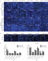
Virus production for the different transfected PK-15 cell groups 24 h after infection. PK-15 cells were transiently transfected with a pCD163-encoding vector in combination with a Siglec-1, Siglec-3, Siglec-5, Siglec-10 or control vector, and 48 h after transfection the cells were treated or not treated with sialidase for 1 h and inoculated with PRRSV LV or MN-184 for 1 h. Twenty-four hours post-infection, the cells were fixed and stained for infection and expression of the different Siglecs and CD163. (a) Immunofluorescence staining of infected cells with mAb 13E2 (against PRRSV nucleocapsid protein; green) [26, 43] and Hoechst 33 342 (nuclei; blue). The absolute number of infected cells for each condition was determined and displayed in the images as the mean ± SEM of three independent experiments. Scale bar: 50 µm. (b) Expression analysis of the different Siglecs using fluorescence microscopy. PK-15 cells were fixed, permeabilized and stained with V5-specific mAb (green) and Hoechst 33 342 (nuclei; blue). The absolute number of transfected cells was determined for each condition and is displayed in the images as the mean ± SEM of three independent experiments. Scale bar: 50 µm. (c) To evaluate virus production, the cell supernatants collected at 24 h p.i. were titrated and the results are displayed in the graphs. The CD163/Siglec double-transfected groups that were significantly different from the CD163 single-transfected group are represented as *P<0.05; **P<0.01 and ***P<0.001.
Fig. 5.
Fig. 5.
Porcine Siglec-10 mediates the endocytosis of PRRSV. (a) Immunofluorescence staining of Siglec-10 in stably transfected PK-15 S10+ cells. PK-15 S10+ cells were fixed with 4 % PF, and the cells were permeabilized (cytoplasmic staining) or not permeablized (surface staining) with 0.1 % Triton X-100 and stained with 1G10 mAb against Siglec-10 (green) and Hoechst 33 342 (nuclei; blue). Scale bar: 25 µm. (b) Attachment and internalization of PRRSV in PK-15 S10+ cells. PK-15 expressing Siglec-10 or normal PK-15 cells were incubated with purified PRRSV LV for 1 h at 4 °C or 37 °C, allowing binding and internalization, respectively. After washing, the cells were fixed and stained with Hoechst 33 342 (nuclei; blue) and mAb 13E2 (PRRSV nucleocapsid protein; green), and analysed by confocal microscopy. Scale bar: 25 µm (c) CHO and PK-15 cells were transiently transfected with wild-type Siglec-10 or the Siglec-10R119E mutant, and a virus internalization assay was performed 48 h post-transfection. Double staining for Siglec-10/Siglec-10R119E (red) and co-localized PRRSV particles (13E2; green) was performed and analysed by confocal microscopy. Scale bar: 25 µm. (d) Quantification of PRRSV internalization in CHO and PK-15 cells for three independent experiments.
Fig. 6.
Fig. 6.
Immunofluorescence staining of Siglec-10/CD21 double-positive cells and Siglec-10/CD163 double-positive cells in tissue sections of the porcine spleen. Immunofluorescence staining of Siglec-10 and CD21 or Siglec-10 and CD163 in tissue sections of the porcine spleen. Tissue samples were sectioned (10 µm) and co-immunostained for Siglec-10 (green) and the markers CD21 (red) or CD163 (red). White dashed lines indicate the border between the B cell-rich area and CD163-rich area. White arrows show CD163+ Siglec-10+ double-positive cells. Scale bar: 25 µm.

Similar articles

Cited by

References

    1. Neumann EJ, Kliebenstein JB, Johnson CD, Mabry JW, Bush EJ, et al. Assessment of the economic impact of porcine reproductive and respiratory syndrome on swine production in the United States. J Am Vet Med Assoc. 2005;227:385–392. doi: 10.2460/javma.2005.227.385. - DOI - PubMed
    1. Meulenberg JJ, Petersen den Besten A, de Kluyver E, van Nieuwstadt A, Wensvoort G, et al. Molecular characterization of Lelystad virus. Vet Microbiol. 1997;55:197–202. doi: 10.1016/S0378-1135(96)01335-1. - DOI - PMC - PubMed
    1. Goldberg TL, Lowe JF, Milburn SM, Firkins LD. Quasispecies variation of porcine reproductive and respiratory syndrome virus during natural infection. Virology. 2003;317:197–207. doi: 10.1016/j.virol.2003.07.009. - DOI - PubMed
    1. Kuhn JH, Lauck M, Bailey AL, Shchetinin AM, Vishnevskaya TV, et al. Reorganization and expansion of the nidoviral family Arteriviridae. Arch Virol. 2016;161:755–768. doi: 10.1007/s00705-015-2672-z. - DOI - PMC - PubMed
    1. Delputte PL, van Gorp H, Favoreel HW, Hoebeke I, Delrue I, et al. Porcine sialoadhesin (CD169/Siglec-1) is an endocytic receptor that allows targeted delivery of toxins and antigens to macrophages. PLoS One. 2011;6:e16827. doi: 10.1371/journal.pone.0016827. - DOI - PMC - PubMed

MeSH terms