

Glaucomatous changes in lamina pores shape within the lamina cribrosa using wide bandwidth, femtosecond mode-locked laser OCT
- PMID: 28742840
- PMCID: PMC5526571
- DOI: 10.1371/journal.pone.0181675
Glaucomatous changes in lamina pores shape within the lamina cribrosa using wide bandwidth, femtosecond mode-locked laser OCT
Abstract
Purpose: The lamina cribrosa (LC) is known to play a critical role in the pathogenesis of glaucoma. Although it has been reported that striae-shaped or slit-shaped lamina pores are more frequent in eyes with primary open angle glaucoma (POAG), this observation is based only on fundus photography. The primary object of this study is to perform layer-by-layer comparisons of the shape of lamina pores within the LC in vivo.
Design: Cross-sectional study.
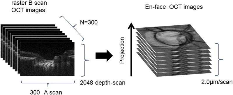
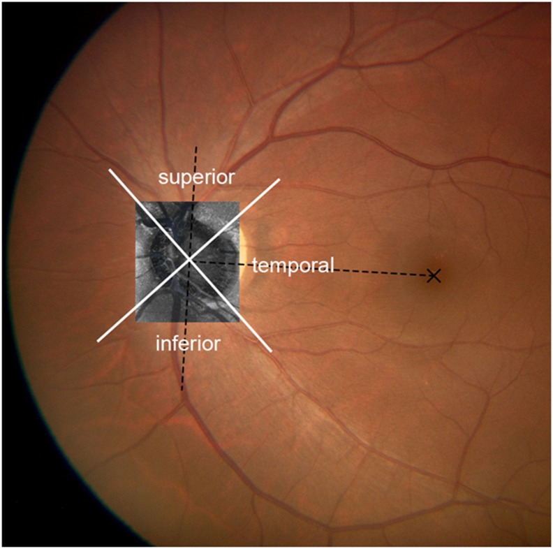
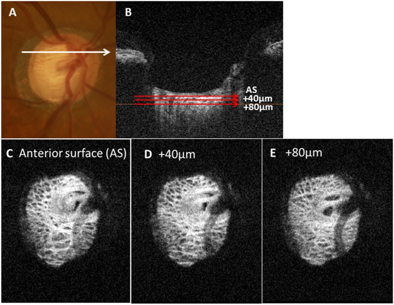
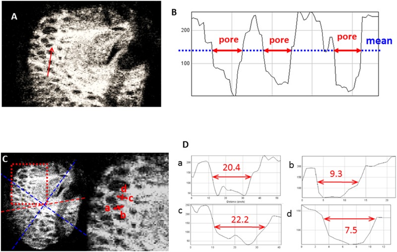
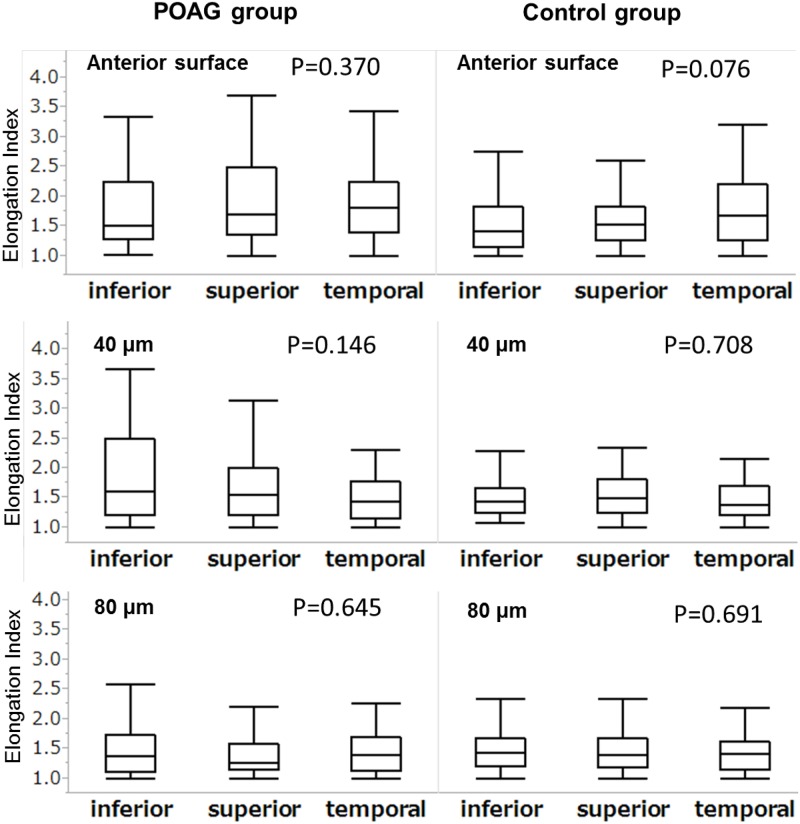
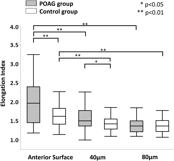
Methods: Optic nerve head B-scans were obtained using custom-made broad-wavelength optical coherence tomography with a mode-locked laser. A total of 300 single B-scans per eye were obtained and three-dimensional images were rendered from these image sequences to obtain 2-μm thin-slice en face images of the LC. Elongation indices (EIs) of the lamina pores were measured from the anterior surface (AS) of the LC to the deeper layers in 40-μm increments.
Results: Thirteen eyes from 10 primary open angle glaucoma (POAG) patients of mean deviation -15.2 (-16.5, -12.9) (median [25,75 percentile]) dB and 10 eyes from 7 normal controls were studied. Although the EI value was not significantly different between the superior, temporal and inferior regions of the LC at any depth level in either group, it was greater at the AS than at the 40 μm and 80 μm depth levels (P < .001) in both groups, and was greater in the POAG group only at the AS and 40 μm depth level (P ≤ .05). After adjustment for age and refraction, the effects of depth and presence of POAG on the EI value remained significant. Also, the severity of glaucoma and depth were significant factors associated with EI in multivariate analysis.
Conclusions: Elongation of lamina pores was significantly more evident at the anterior surface and the 40-μm depth level of the LC in POAG eyes than in normal eyes, suggesting that nerve fiber bundles passing through the LC were under greater stress in the anterior layers of the LC.
Conflict of interest statement
Figures
References
-
- Radius RL, Gonzales M. Anatomy of the lamina cribrosa in human eyes. Arch Ophthalmol. 1981;99(12):2159–62. . - PubMed
-
- Quigley HA, Addicks EM, Green WR, Maumenee AE. Optic nerve damage in human glaucoma. II. The site of injury and susceptibility to damage. Arch Ophthalmol. 1981;99(4):635–49. . - PubMed
-
- Roberts MD, Sigal IA, Liang Y, Burgoyne CF, Downs JC. Changes in the biomechanical response of the optic nerve head in early experimental glaucoma. Invest Ophthalmol Vis Sci. 2010;51(11):5675–84. Epub 2010/06/12. doi: 10.1167/iovs.10-5411 . - DOI - PMC - PubMed
-
- Jonas JB, Mardin CY, Schlotzer-Schrehardt U, Naumann GO. Morphometry of the human lamina cribrosa surface. Invest Ophthalmol Vis Sci. 1991;32(2):401–5. Epub 1991/02/01. . - PubMed
MeSH terms
LinkOut - more resources
Full Text Sources
Other Literature Sources
Research Materials
Miscellaneous
