. 2017 Jul 25;12(7):e0181822.
doi: 10.1371/journal.pone.0181822. eCollection 2017.

In vitro and in vivo anti-inflammatory active copper(II)-lawsone complexes

Affiliations

In vitro and in vivo anti-inflammatory active copper(II)-lawsone complexes

Ján Vančo et al. PLoS One. .

Abstract

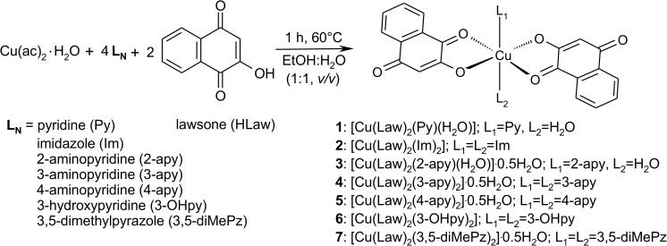
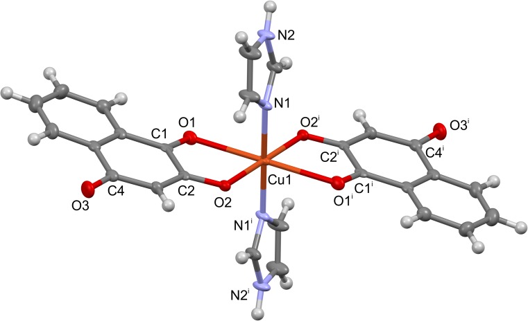
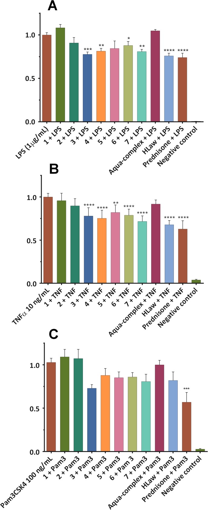
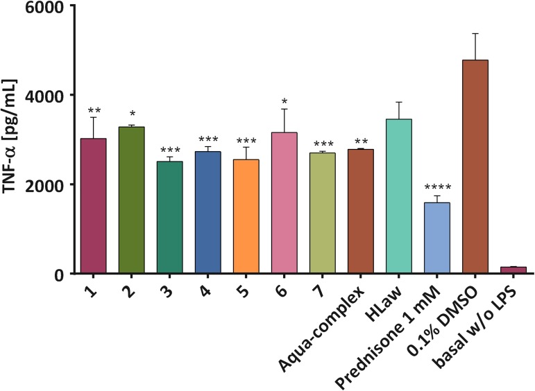
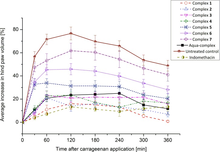
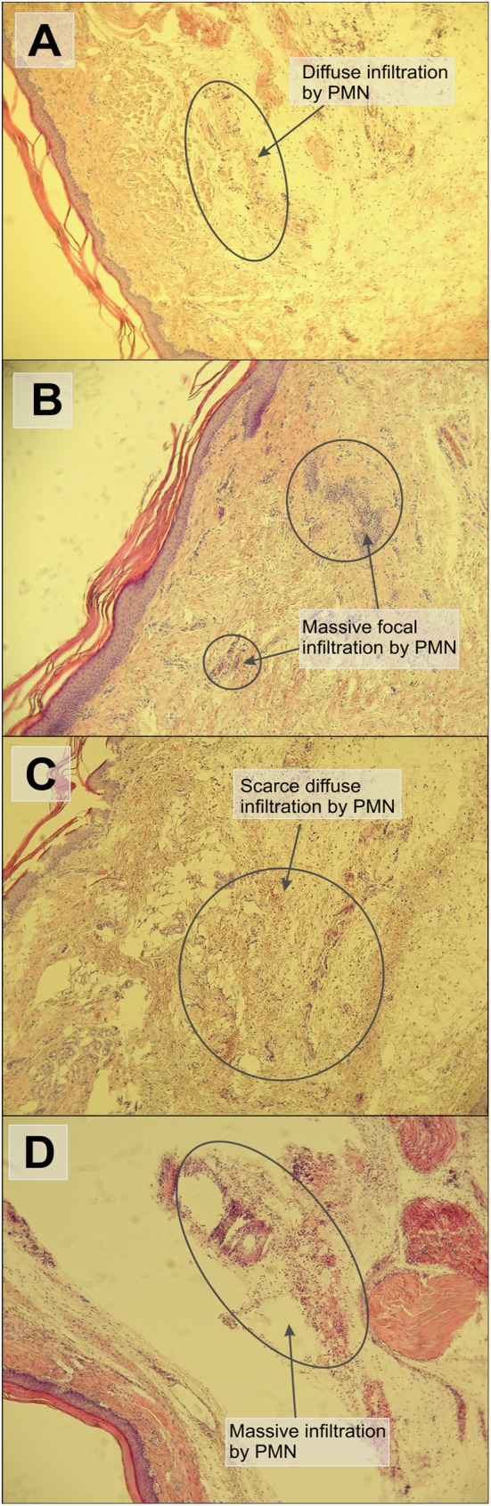
We report in vitro and in vivo anti-inflammatory activities of a series of copper(II)-lawsone complexes of the general composition [Cu(Law)2(LN)x(H2O)(2-x)]·yH2O; where HLaw = 2-hydroxy-1,4-naphthoquinone, x = 1 when LN = pyridine (1) and 2-aminopyridine (3) and x = 2 when LN = imidazole (2), 3-aminopyridine (4), 4-aminopyridine (5), 3-hydroxypyridine (6), and 3,5-dimethylpyrazole (7). The compounds were thoroughly characterized by physical techniques, including single crystal X-ray analysis of complex 2. Some of the complexes showed the ability to suppress significantly the activation of nuclear factor κB (NF-κB) both by lipopolysaccharide (LPS) and TNF-alpha (complexes 3-7 at 100 nM level) in the similar manner as the reference drug prednisone (at 1 μM level). On the other hand, all the complexes 1-7 decreased significantly the levels of the secreted TNF-alpha after the LPS activation of THP-1 cells, thus showing the anti-inflammatory potential via both NF-κB moderation and by other mechanisms, such as influence on TNF-alpha transcription and/or translation and/or secretion. In addition, a strong intracellular pro-oxidative effect of all the complexes has been found at 100 nM dose in vitro. The ability to suppress the inflammatory response, caused by the subcutaneous application of λ-carrageenan, has been determined by in vivo testing in hind-paw edema model on rats. The most active complexes 1-3 (applied in a dose corresponding to 40 μmol Cu/kg), diminished the formation of edema simalarly as the reference drug indomethacine (applied in 10 mg/kg dose). The overall effect of the complexes, dominantly 1-3, shows similarity to anti-inflammatory drug benoxaprofen, known to induce intracellular pro-oxidative effects.


Conflict of interest statement

Competing Interests: The authors have declared that no competing interests exist.

Figures

Fig 1
Fig 1
The general formula of 1,4-naphthoquinone showing most usual positions of substitutions, as indicated by arrows (A), and the formulas of selected 1,4-naphthoquinone derivatives isolated from natural sources: lawsone (B), juglone (C), and lapachol (D).
Fig 2
Fig 2. The general pathway of the preparation of complexes 1–7.
Fig 3
Fig 3. The result of temperature-dependent SQUID magnetometry measurements for complex 5.
The curves represent the best fit of experimental data according to the Curie-Weiss law (the best-fit parameters are giso = 2.18, C = 5.585×10⁻⁶ m3.mol-1, and θ = -0.181 K with a temperature-independent paramagnetism term aTIP = 3.40×10⁻⁸ m3.mol-1). C0 = 4.7141997×10⁻⁶ m3.mol-1.
Fig 4
Fig 4. The molecular structure of complex 2.
Symmetry code used: i = -x+1,-y,-z+1.
Fig 5
Fig 5. Effect of complexes 1–7 on the NF-κB activity.
THP1-XBlue™-MD2-CD14 cell line were pre-treated with the complexes (100 nM, dissolved in PBS), [Cu(Law)2(H2O)2]·0.5H2O, HLaw and prednisone (1 μM, dissolved in DMSO) for 1 h. Subsequently, LPS (1 μg/mL) (A) or TNF-α (10 ng/mL) (B) or Pam3CSK4 (100 ng/mL) (C) was added [except for the control cells (basal)] to trigger the activation of NF-κB. After 24 h, the activity of NF-κB was evaluated based on the amount of the secreted alkaline phosphatase measured spectrophotometrically. The results are expressed as the mean ± S.E.M. for six independent experiments. ** Indicates a significant difference in comparison with the vehicle-treated cells p < 0.01, and **** indicates a significant difference in comparison with the vehicle-treated cells p < 0.0001.
Fig 6
Fig 6. The pro-oxidative effect of complexes 1–7, reference aqua-complex and lawsone determined by dichlorofluorescein method.
THP1-XBlue™-MD2-CD14 cell line were treated with the complexes, or standard antioxidant Trolox® at the final concentration of 100 nM for 1 h (left diagram) and 24 h (right diagram). The data represent the relative increase of fluorescence intensity after the addition of DCFH-DA in comparison with the untreated control. The results are expressed as the mean values ± S.E.M. for three independent experiments.
Fig 7
Fig 7. The effect of the tested complexes on the secretion of TNF-α.
THP-1 macrophages were pre-treated with the compounds 17, [Cu(Law)2(H2O)2]·0.5H2O (aqua-complex), HLaw (100 nM, dissolved in PBS) and prednisone (1 μM, dissolved in DMSO) for 1 h. Subsequently, LPS (1 μg/mL) was added [except for the control cells (basal)] to trigger the secretion of the pro-inflammatory cytokine TNF-α. After 24 h, the amount of the secreted TNF-α was evaluated by ELISA. The results are expressed as the mean values ± S.E.M. for three independent experiments. * Indicates a significant difference in comparison with the vehicle-treated cells p < 0.05, ** indicates a significant difference in comparison with the vehicle-treated cells p < 0.01, *** indicates a significant difference in comparison with the vehicle-treated cells p < 0.001, and **** indicates a significant difference in comparison with the vehicle-treated cells p < 0.0001.
Fig 8
Fig 8. The time-resolved changes in the average volume of the hind paws of rats.
The values are expressed as average values ± S.E.M. calculated for 7 animals in each experimental group. The copper(II) complexes were applied i.p. at the dose corresponding to 40 mmol Cu/kg (ca. 20 mg/kg), and indomethacin at the i.p. dose of 5 mg/kg.
Fig 9
Fig 9. Histological evaluation of inflammatory response in tissue sections of the hind paw, stained by hematoxylin/eosin (40x magnification).
The section of plantar tissues from the groups pretreated with complex 1 (A) or indomethacin (C) with the weak inflammatory response in the hypodermis with scarce PMN infiltrate. The tissue sections from the group exposed to complex 7 (B) and 10% DMF solution (control, D) with the strong inflammatory reaction in the hypodermis with massive focal PMN infiltrate.

References

    1. Pradhan R, Dandawate P, Vyas A, Padhye S, Biersack B, Schobert R, et al. From Body Art to Anticancer Activities: Perspectives on Medicinal Properties of Henna, Curr. Drug Targets 2012; 13: 1777–1798. - PubMed
    1. Singh DK, Luqman S, Mathur AK. Lawsonia inermis L.–A commercially important primaeval dying and medicinal plant with diverse pharmacological activity: A review. Ind. Crop. Prod. 2015; 65: 269–286.
    1. Biradar S, Veeresh B. Protective effect of lawsone on L-Arginine induced acute pancreatitis in rats. Indian J. Exp. Biol. 2013; 51: 256–261. - PubMed
    1. Ali BH, Bashir AK, Tanira MOM. Anti-inflammatory, antipyretic, and analgesic effects of Lawsonia inermis L (Henna) in rats. Pharmacology 1995; 51: 356–363. - PubMed
    1. Marzin D, Kirkland D. 2-Hydroxy-1,4-naphthoquinone, the natural dye of Henna, is non-genotoxic in the mouse bone marrow micronucleus test and does not produce oxidative DNA damage in Chinese hamster ovary cells. Mutat. Res. 2004; 560: 41–47. doi: 10.1016/j.mrgentox.2004.02.004 - DOI - PubMed

LinkOut - more resources