. 2017 Jul 25;318(4):360-370.
doi: 10.1001/jama.2017.8334.

Clinicopathological Evaluation of Chronic Traumatic Encephalopathy in Players of American Football

Affiliations

Clinicopathological Evaluation of Chronic Traumatic Encephalopathy in Players of American Football

Jesse Mez et al. JAMA. .

Abstract

Importance: Players of American football may be at increased risk of long-term neurological conditions, particularly chronic traumatic encephalopathy (CTE).

Objective: To determine the neuropathological and clinical features of deceased football players with CTE.

Design, setting, and participants: Case series of 202 football players whose brains were donated for research. Neuropathological evaluations and retrospective telephone clinical assessments (including head trauma history) with informants were performed blinded. Online questionnaires ascertained athletic and military history.

Exposures: Participation in American football at any level of play.

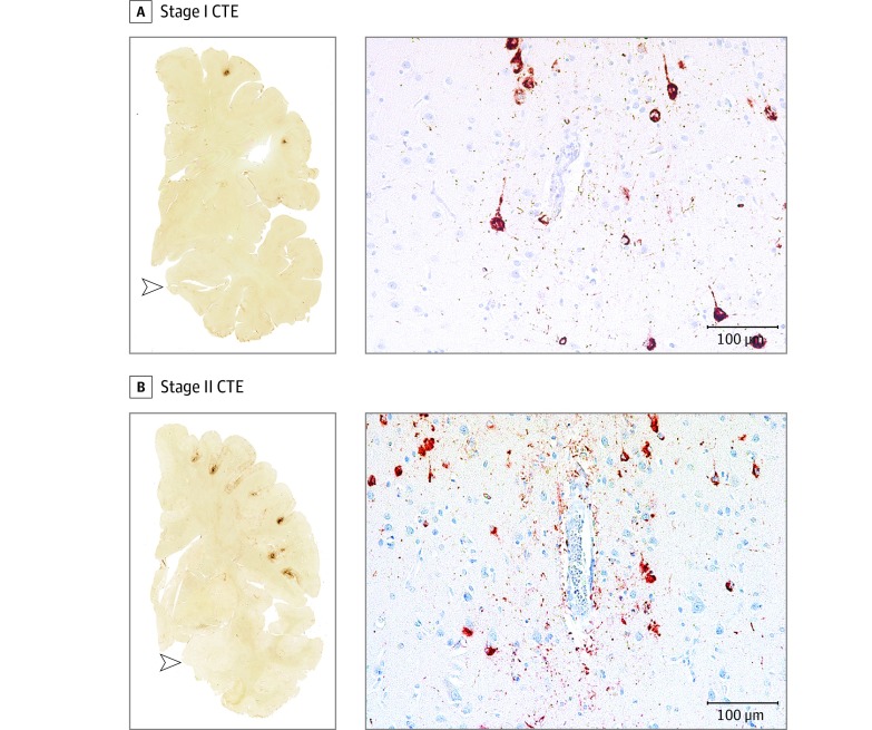
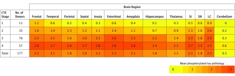
Main outcomes and measures: Neuropathological diagnoses of neurodegenerative diseases, including CTE, based on defined diagnostic criteria; CTE neuropathological severity (stages I to IV or dichotomized into mild [stages I and II] and severe [stages III and IV]); informant-reported athletic history and, for players who died in 2014 or later, clinical presentation, including behavior, mood, and cognitive symptoms and dementia.

Results: Among 202 deceased former football players (median age at death, 66 years [interquartile range, 47-76 years]), CTE was neuropathologically diagnosed in 177 players (87%; median age at death, 67 years [interquartile range, 52-77 years]; mean years of football participation, 15.1 [SD, 5.2]), including 0 of 2 pre-high school, 3 of 14 high school (21%), 48 of 53 college (91%), 9 of 14 semiprofessional (64%), 7 of 8 Canadian Football League (88%), and 110 of 111 National Football League (99%) players. Neuropathological severity of CTE was distributed across the highest level of play, with all 3 former high school players having mild pathology and the majority of former college (27 [56%]), semiprofessional (5 [56%]), and professional (101 [86%]) players having severe pathology. Among 27 participants with mild CTE pathology, 26 (96%) had behavioral or mood symptoms or both, 23 (85%) had cognitive symptoms, and 9 (33%) had signs of dementia. Among 84 participants with severe CTE pathology, 75 (89%) had behavioral or mood symptoms or both, 80 (95%) had cognitive symptoms, and 71 (85%) had signs of dementia.

Conclusions and relevance: In a convenience sample of deceased football players who donated their brains for research, a high proportion had neuropathological evidence of CTE, suggesting that CTE may be related to prior participation in football.


Conflict of interest statement

Conflict of Interest Disclosures: All authors have completed and submitted the ICMJE Form for Disclosure of Potential Conflicts of Interest. Dr Nowinski reported that he receives travel reimbursements for various unpaid advisory roles from the NFL Players’ Association, Major League Lacrosse, World Wrestling Entertainment (WWE), National Collegiate Athletic Association (NCAA), and the Ivy League; receives royalties from the publication of his book Head Games: The Global Concussion Crisis, published by Head Games The Film; served as a consultant for MC10 Inc as recently as 2013; serves as chief executive officer of the Concussion Legacy Foundation; and receives speaking honoraria and travel reimbursements for educational lectures. Ms Baugh reported that she receives research funding through the NCAA and the Harvard Football Players Health Study, which is funded by the NFL Players’ Association. Dr Cantu reported that he receives compensation from the NFL as senior advisor to its Head, Neck and Spine Committee, from the National Operating Committee on Standards for Athletic Equipment as chair of its Scientific Advisory Committee and from the Concussion Legacy Foundation as cofounder and medical director for some talks given and receives royalties from Houghton Mifflin Harcourt and compensation from expert legal opinion. Dr Stern reported that he has received research funding from the NFL, the NFL Players’ Association, and Avid Radiopharmaceuticals Inc; is a member of the Mackey-White Committee of the NFL Players’ Association; is a paid consultant to Amarantus BioScience Holdings Inc, Avanir Pharmaceuticals Inc, and Biogen; and receives royalties for published neuropsychological tests from Psychological Assessment Resources Inc and compensation from expert legal opinion. Dr McKee reported that she has received funding from the NFL and WWE and is a member of the Mackey-White Committee of the NFL Players’ Association.

Figures

Figure 1.
Figure 1.. Representative Images of Phosphorylated Tau Pathology at CTE Pathological Stages I and II
CTE indicates chronic traumatic encephalopathy; NFT, neurofibrillary tangle; ptau, phosphorylated tau. For all images, 10-µm paraffin-embedded tissue sections were immunostained with microscopic mouse monoclonal antibody for phosphorylated tau (AT8) (Pierce Endogen). Positive ptau immunostaining appears dark red, hematoxylin counterstain; calibration bar indicates 100 µm. Stage I CTE is characterized by 1 or 2 isolated perivascular epicenters of ptau NFTs and neurites (ie, CTE lesions) at the depths of the cortical sulci. In stage II, 3 or more cortical CTE lesions are found. All hemispheric tissue section images are 50-µm sections immunostained with mouse monoclonal antibody CP-13, directed against phosphoserine 202 of tau (courtesy of Peter Davies, PhD, Feinstein Institute for Medical Research; 1:200); this is considered to be an early site of tau phosphorylation in NFT formation. Positive ptau immunostaining appears dark brown. A, Former college football player with stage I CTE. Two perivascular ptau CTE lesions are evident at sulcal depths of the frontal cortex; there is no neurofibrillary degeneration in the medial temporal lobe (open arrowhead). Perivascular CTE lesion: neurofibrillary tangles and dot-like and threadlike neurites encircle a small blood vessel. B, Former NFL player with stage II CTE. There are multiple perivascular ptau CTE lesions at depths of sulci of the frontal cortex; there is no neurofibrillary degeneration in the medial temporal lobe (open arrowhead). Perivascular CTE lesion: a cluster of NFTs and large dot-like and threadlike neurites surround a small blood vessel.
Figure 2.
Figure 2.. Representative Images of Phosphorylated Tau Pathology at CTE Pathological Stages III and IV
CTE indicates chronic traumatic encephalopathy; NFT, neurofibrillary tangle; ptau, phosphorylated tau. For all images, 10-µm paraffin-embedded tissue sections were immunostained with microscopic mouse monoclonal antibody for phosphorylated tau (AT8) (Pierce Endogen). Positive ptau immunostaining appears dark red, hematoxylin counterstain; calibration bar indicates 100 µm. In stage III CTE, multiple CTE lesions and diffuse neurofibrillary degeneration of the medial temporal lobe are found. In stage IV CTE, CTE lesions and NFTs are widely distributed throughout the cerebral cortex, diencephalon, and brain stem. All hemispheric tissue section images are 50-µm sections immunostained with mouse monoclonal antibody CP-13, directed against phosphoserine 202 of tau (courtesy of Peter Davies, PhD, Feinstein Institute for Medical Research; 1:200); this is considered to be an early site of tau phosphorylation in NFT formation. Positive ptau immunostaining appears dark brown. A, Former NFL player with stage III CTE. There are multiple large CTE lesions in the frontal cortex and insula; there is diffuse neurofibrillary degeneration of hippocampus and entorhinal cortex (black arrowhead). Perivascular CTE lesion: a dense collection of NFTs and large dot-like and threadlike neurites enclose several small blood vessels. B, Former NFL player with stage IV CTE. There are large, confluent CTE lesions in the frontal, temporal, and insular cortices and there is diffuse neurofibrillary degeneration of the amygdala and entorhinal cortex (black arrowhead). Perivascular CTE lesion: a large accumulation of NFTs, many of them ghost tangles, encompass several small blood vessels.
Figure 3.
Figure 3.. Phosphorylated Tau Pathology for Each Brain Region by CTE Neuropathological Stage
CTE indicates chronic traumatic encephalopathy; NFT: neurofibrillary tangle, SI: substantia innominata, SN: substantia nigra; LC: locus coeruleus. Cerebellum indicates dentate nucleus of the cerebellum. In each region, 0 = no NFTs (yellow); 1 = 1 NFT per 20× field (orange); 2 = 2 to 3 NFTs per 20× field (amber); and 3 = ≥4 NFTs per 20× field (red). The color scale is based on the distribution of all values, not by each individual stage. Values represent means of phosphorylated tau pathology among participants in each stage.

Comment in

References

    1. Corsellis JA, Bruton CJ, Freeman-Browne D. The aftermath of boxing. Psychol Med. 1973;3(3):270-303. - PubMed
    1. Hof PR, Knabe R, Bovier P, Bouras C. Neuropathological observations in a case of autism presenting with self-injury behavior. Acta Neuropathol. 1991;82(4):321-326. - PubMed
    1. Geddes JF, Vowles GH, Nicoll JA, Révész T. Neuronal cytoskeletal changes are an early consequence of repetitive head injury. Acta Neuropathol. 1999;98(2):171-178. - PubMed
    1. Omalu BI, DeKosky ST, Minster RL, Kamboh MI, Hamilton RL, Wecht CH. Chronic traumatic encephalopathy in a National Football League player. Neurosurgery. 2005;57(1):128-134. - PubMed
    1. Omalu BI, DeKosky ST, Hamilton RL, et al. . Chronic traumatic encephalopathy in a National Football League player: part II. Neurosurgery. 2006;59(5):1086-1092. - PubMed

Publication types

MeSH terms