. 2017 Jul 25;17(1):516.
doi: 10.1186/s12879-017-2621-4.

Magnetic hyperthermia enhance the treatment efficacy of peri-implant osteomyelitis

Affiliations

Magnetic hyperthermia enhance the treatment efficacy of peri-implant osteomyelitis

Chih-Hsiang Fang et al. BMC Infect Dis. .

Abstract

Background: When bacteria colony persist within a biofilm, suitable drugs are not yet available for the eradication of biofilm-producing bacteria. The aim of this study is to study the effect of magnetic nano-particles-induced hyperthermia on destroying biofilm and promoting bactericidal effects of antibiotics in the treatment of osteomyelitis.

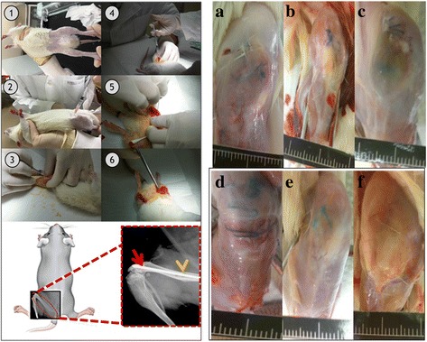
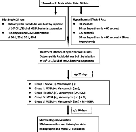
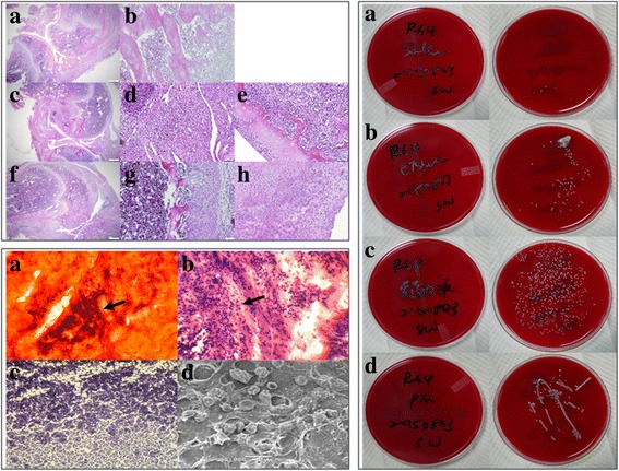
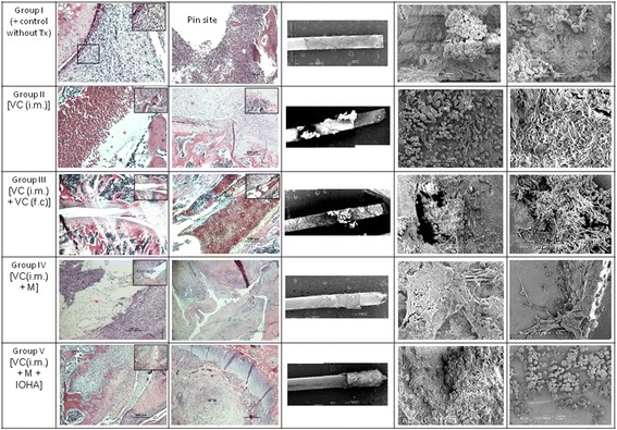
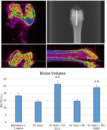
Methods: Sixty 12-weeks-old male Wistar rats were used. A metallic 18G needle was implanted into the bone marrow cavity of distal femur after the injection of Methicillin-sensitive Staphylococcus aureus (MSSA). All animals were divided into 5 different treatment modalities. The microbiological evaluation, scanning electron microscope examination, radiographic examination and then micro-CT evaluation of peri-implant bone resorption were analyzed.

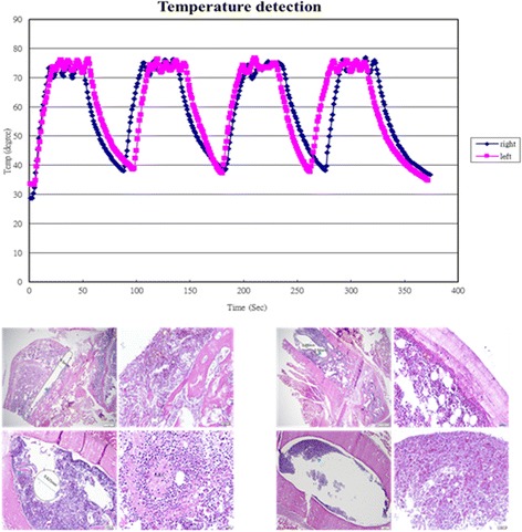
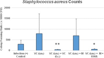
Results: The pathomorphological characteristics of biofilm formation were completed after 40-days induction of osteomyelitis. The inserted implants can be heated upto 75 °C by magnetic heating without any significant thermal damage on the surrounding tissue. We also demonstrated that systemic administration of vancomycin [VC (i.m.)] could not eradicate the bacteria; but, local administration of vancomycin into the femoral canal and the presence of magnetic nanoparticles hyperthermia did enhance the eradication of bacteria in a biofilm-based colony. In these two groups, the percent bone volume (BV/TV: %) was significantly higher than that of the positive control.

Conclusions: For the treatment of chronic osteomyelitis, we developed a new modality to improve antibiotic efficacy; the protection effect of biofilms on bacteria could be destroyed by magnetic nanoparticles-induced hyperthermia and therapeutic effect of systemic antibiotics could be enhanced.

Keywords: Biofilm; Hyperthermia; Magnetic nanoparticle; Peri-implant osteomyelitis.


Conflict of interest statement

Ethics approval and consent to participate

In this manuscripts no human participants, human data or human tissue are included.

The experimental protocol was approved by the Institutional Animal Care and Use Committee of Medical College, National Taiwan University (Taipei, Taiwan).

Consent for publication

Not applicable.

Competing interests

The authors declare that there is no benefits in any form have been or will be received from a commercial party related directly or indirectly to the subject of this manuscript.

Publisher’s Note

Springer Nature remains neutral with regard to jurisdictional claims in published maps and institutional affiliations.

Figures

Fig. 1
Fig. 1
Sequence of surgical procedures and X-ray image and Animal models for induction of osteomyelitis. Left: Sequence of surgical procedures and X-ray image. Osteomyelitis animal models used 18G needle as implant and injecting methicillin-sensitive Staphylococcus aureus suspension into bone marrow canal as an infection model. The X-ray image showed that needle completely inserted into femur of rats. Right: Animal models for induction of osteomyelitis. a Uninfected group (left leg); (b) Uninfected group (right leg); (c) Osteomyelitis group (left leg): 10 days after induction of infection; (d) Osteomyelitis group (right leg): 10 days after induction of infection; (e) Osteomyelitis group (left leg): 40 days after induction of infection; (f) Osteomyelitis group (right leg): 40 days after induction of infection
Fig. 2
Fig. 2
Flow Chart of experimental design
Fig. 3
Fig. 3
Transmission Electron Microscope (TEM) examination of Fe3O4 magnetic nanoparticles. Upper: TEM of F3O4 Nanoparticles: Effect of different amounts of stabilizer and dispersant. a 80 μL, b 160 μL, and c 400 μL surfactant. Lower: TEM of F3O4 Nanoparticles: Effect of different temperature on the formation F3O4 Nanoparticles. Injection temperature: a 260 °C, b 230 °C, and c 200 °C
Fig. 4
Fig. 4
Characteristics of Fe3O4 nanoparticles. a The XRD pattern of the Fe3O4 nanoparticles. b Room-temperature magnetization curves for FO111019-P and FO111019-h. Magnetic hysteresis curves measured at room temperature for the samples prepared by a hydrothermal process (square: FO111019-h, dot: FO111019-p) showed the super-paramagnetic characteristics. c The crystal structure of Fe3O4 nanoparticles prepared at the injection temperature of 290 °C (TEM). The average diameter of particles is 16 nm
Fig. 5
Fig. 5
Tissue damage after thermotherapy (H & E staining). Left: The needle at 75 °C for 90 s by magnetic heating (each 30 s heating with a rest for 1 min), Right: The needle at 75 °C for 120 s by magnetic heating (each 30 s heating with a rest for 1 min). There were no significant thermal damage noted in these two testing conditions
Fig. 6
Fig. 6
Histological, histopathological staining and microbiology assessment from osteomyelitis rats after 40 days infection. Left upper: Histological sections with H&E staining. Upper: Uninfected group, magnification a 40 × and b 1000 ×; Middle: Osteomyelitis 10 days after induction of infection, magnification c 40 ×, d and e 1000 ×; Lower: Osteomyelitis 40 days after induction of infection, magnification f 40 ×, g and h 1000 ×. In this figure, uninfected group possesses normal trabecular patterns of cancellous bone (upper: a and b); while in the acute suppurative changes of osteomyelitis, there is devitalized lamellar bone with scalloped edges and absence of stainable osteocytes and osteoblasts, edema and granulocytic infiltration of surrounding tissues are obvious (middle: c-e). In the chronic osteomyelitis, there is the irregular fragment of devitalized bone surrounded by dense fibrous tissue heavily infiltrated by plasma cells, lymphocytes, and only a few granulocytes (lower: f-h). Left lower: Histopathological staining. a Cango red staining, b H&E staining, c Gram staining and d SEM morphology, respectively. As shown here, GRAM-staining revealed the presence of bacteria within the defect site as Gram (+) Staphylococcus aureus bacteria could be detected (b and c). Cango red staining (a) and SEM image (d) showed the biofilm forming completed after 40 days induction of osteomyelitis. Magnification of pictures are 1000 ×. Right: Microbiology assessment from osteomyelitis rats after 40 days infection. Specimens streaked onto agar plate for: a Sterilized PBS as control group; b Tissue from the affected area; c Intramedullary liquid from the affected femur; d Implants from the affected animals. All smears taken from the experimental sites showed characteristic accretion of S. aureus infection. In the control group, there is few characteristic CFUs on the agar plate (a). On the other hand, tissue from the affected area, intramedullary liquid from the infected femur and the implants from the infected animals all showed characteristic growth of CFUs on agar plates (b-d)
Fig. 7
Fig. 7
Histological and scanning electron microscope examination of different treatment modalities for osteomyelitis rats after 40 days infection. After inoculation of Staphylococcus aureus, tissue necrosis with active inflammation and even biofilm formation was clearly demonstrated in all groups; while, the biofilm formation were found both the at the surface of the inserted pin and the inner surface of bony trabeculae. Group I: osteomyelitis positive control without treatment; Group II: osteomyelitis treated with intramuscular injection of vancomycin [VC (i.m.)]; Group III: osteomyelitis treated with both intramuscular and femur cavity injection of vancomycin [VC (i.m.) + VC (f.c)]; Group IV: osteomyelitis treated with both intramuscular injection of vancomycin and magnetic particles [VC(i.m.) + M]; Group V: osteomyelitis treated with intramuscular injection of vancomycin and magnetic particles hyperthermia [VC(i.m.) + M + IOHA]. Animals from each group were sacrificed by overdose of pentobarbital at 40 days after surgery. *(red): biofilm-like ECM; **(black): necrosis with inflammation; ***(green): inflammation
Fig. 8
Fig. 8
Colony-forming unit of implants and the affected area under different treatment modalities. Systemic administration of vancomycin [VC (i.m.)] did not show any better effect on eradication of the bacteria; similar event observed in the presence of magnetic nanoparticles [VC (i.m.) + M]. Local administration of vancimycin into the femoral canal [VC (i.m.) + VC (f.c)] did enhance the eradication of bacteria; similar event observed in the presence of magnetic nanoparticles when magnetic field was applied [VC (i.m.) + M + IOHA]
Fig. 9
Fig. 9
Change of relative bone volume at the affected area under different treatment modalities. Upper: Micro-CT (Computed Tomography) evaluation and radiographic examination. Lower: Systemic administration of vancomycin [VC (i.m.)] with/or without the presence of magnetic nanoparticles [VC (i.m.) + M] did not affect the bone volume change under peri-implent osteomyelitis condition; but, percent of bone volume (BV/TV) was significantly higher in the group of local administration of vancomycin into the femoral canal [VC (i.m.) + VC (f.c)] and the group when magnetic field was applied in the presence of magnetic nanoparticles [VC (i.m.) + M + IOHA]

References

    1. Gustilo RB, Merkow RL, Templeman D. The management of open fractures. J Bone Joint Surg Am. 1990;72(2):299–304. doi: 10.2106/00004623-199072020-00023. - DOI - PubMed
    1. Walter G, Kemmerer M, Kappler C, Hoffmann R. Treatment algorithms for chronic osteomyelitis. Dtsch Arztebl Int. 2012;109(14):257–264. - PMC - PubMed
    1. Lew DP, Waldvogel FA. Osteomyelitis. Lancet. 2004;364(9431):369–379. doi: 10.1016/S0140-6736(04)16727-5. - DOI - PubMed
    1. Costerton JW. Biofilm theory can guide the treatment of device-related orthopaedic infections. Clin Orthop Relat Res. 2005;437:7–11. doi: 10.1097/00003086-200508000-00003. - DOI - PubMed
    1. Marais LC, Ferreira N, Aldous C, Le Roux TL. The outcome of treatment of chronic osteomyelitis according to an integrated approach. Strategies Trauma Limb Reconstr. 2016;11(2):135–142. doi: 10.1007/s11751-016-0259-1. - DOI - PMC - PubMed

MeSH terms