. 2017 Jul 25;12(1):55.
doi: 10.1186/s13024-017-0196-6.

The lysosomal protein cathepsin L is a progranulin protease

Affiliations

The lysosomal protein cathepsin L is a progranulin protease

Chris W Lee et al. Mol Neurodegener. .

Abstract

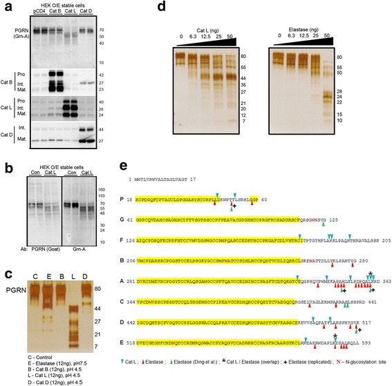
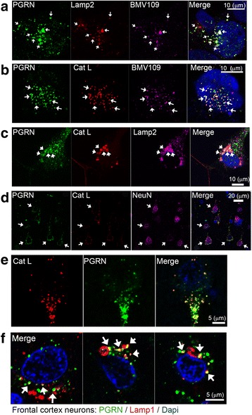
Haploinsufficiency of GRN, the gene encoding progranulin (PGRN), causes frontotemporal lobar degeneration (FTLD), the second most common cause of early-onset dementia. Receptor-mediated lysosomal targeting has been shown to regulate brain PGRN levels, and complete deficiency of PGRN is a direct cause of neuronal ceroid lipofuscinosis (NCL), a lysosomal storage disease. Here we show that the lysosomal cysteine protease cathepsin L (Cat L) can mediate the proteolytic cleavage of intracellular PGRN into poly-granulin and granulin fragments. Further, PGRN and Cat L co-localize in lysosomes of HEK293 cells, iPSC-derived neurons and human cortical neurons from human postmortem tissue. These data identify Cat L as a key intracellular lysosomal PGRN protease, and provides an intriguing new link between lysosomal dysfunction and FTLD.

Keywords: Cathepsin L; Frontotemporal lobar degeneration; Lysosome; Neuronal ceroid lipofuscinosis; Neutrophil elastase; Progranulin.


Conflict of interest statement

Ethics approval and consent to participate

Not applicable.

Consent for publication

All authors have reviewed the final manuscript and consent to publication.

Competing interests

The authors declare that they have no competing interests.

Publisher’s Note

Springer Nature remains neutral with regard to jurisdictional claims in published maps and institutional affiliations.

Figures

Fig. 1
Fig. 1
Cathepsin L proteolytically processes intracellular PGRN, and in a manner distinct from elastase. a HEK293 stable cell lines stably overexpressing pCDNA4 (pCD4), Cat B, Cat L and Cat D were analyzed for levels of PGRN and its fragments by western blot using Grn-A antibody. The expression levels of different forms (pro-, intermediate- and mature- [40, 41]) of Cat B, Cat L and Cat D were validated in the stable cell lines. b A second, full-length PGRN polyclonal goat antibody was applied to detect PGRN and its proteolytic fragments in HEK-Cat L cells in comparison to HEK-pCDNA4 cells (Con). c Recombinant PGRN was incubated with same amount of elastase (E), Cat B (B), Cat L (L) or Cat D (D) optimal reaction conditions. Cat L, but not Cat B or Cat D, processed PGRN into protein fragments after the reaction. d Recombinant PGRN protein products were detected after the addition of increasing concentrations of recombinant Cat L (left panel) or elastase (right panel). e Cleavage sites used by Cat L and elastase on PGRN were mapped by LC/MS analysis of the small peptides produced from the in vitro proteolytic reaction. All detected peptides are listed in Tables 1 and 2. Annotated MS/MS spectra of the identified granulin peptides were included in Additional file 1: Figure S2. Cat L and elastase cleavage sites are labeled by blue triangle and red triangle, respectively. Common cleavage sites are highlighted by the asterisk sign. Previously published elastase cleavage sites are labeled by green triangle. Elastase cleavage sites replicated from the current study are highlighted by plus sign. N-glycosylation sites were highlighted in red
Fig. 2
Fig. 2
Cellular PGRN co-localize with Cat L and active cysteine cathepsins in lysosomes. a Immunofluorescence (IF) analysis of HEK293 cells showing the localization of PGRN (green), Lamp2 (red) and active cysteine cathepsins (BMV109 activity probe; violet). Nuclei in merged images were visualized by Hoechst 33,342 staining. b IF analysis of PGRN (green), Cat L (red) and BMV109 (violet) in HEK293 cells. c IF analysis of human wild-type iPSC-neurons showing the localization of PGRN (green), Cat L (red), and Lamp2 (violet). d IF analysis of neuronal cells in the frontal cortex of a sporadic FTLD patient showing the localization of PGRN (green), Cat L (red), and NeuN (violet). e High magnification IF analysis of Cat L (red) and PGRN (green) in a cortical neuron from the frontal cortex. f High magnification IF analysis of 3 representative cortical neurons in the frontal cortex of a sporadic FTLD patient showing the localization of PGRN (green), Lamp1 (red), and the nucleus (DAPI; blue). The white arrows show triple co-localization (ac) or co-localization of PGRN and Cat L in NeuN position cells (d)

References

    1. Ng AS, Rademakers R, Miller BL. Frontotemporal dementia: a bridge between dementia and neuromuscular disease. Ann N Y Acad Sci. 2015;1338:71–93. doi: 10.1111/nyas.12638. - DOI - PMC - PubMed
    1. Shankaran SS, Capell A, Hruscha AT, Fellerer K, Neumann M, Schmid B, Haass C. Missense mutations in the progranulin gene linked to frontotemporal lobar degeneration with ubiquitin-immunoreactive inclusions reduce progranulin production and secretion. J Biol Chem. 2008;283:1744–1753. doi: 10.1074/jbc.M705115200. - DOI - PubMed
    1. Gass J, Cannon A, Mackenzie IR, Boeve B, Baker M, Adamson J, Crook R, Melquist S, Kuntz K, Petersen R, et al. Mutations in progranulin are a major cause of ubiquitin-positive frontotemporal lobar degeneration. Hum Mol Genet. 2006;15:2988–3001. doi: 10.1093/hmg/ddl241. - DOI - PubMed
    1. Baker M, Mackenzie IR, Pickering-Brown SM, Gass J, Rademakers R, Lindholm C, Snowden J, Adamson J, Sadovnick AD, Rollinson S, et al. Mutations in progranulin cause tau-negative frontotemporal dementia linked to chromosome 17. Nature. 2006;442:916–919. doi: 10.1038/nature05016. - DOI - PubMed
    1. Cruts M, Gijselinck I, van der Zee J, Engelborghs S, Wils H, Pirici D, Rademakers R, Vandenberghe R, Dermaut B, Martin JJ, et al. Null mutations in progranulin cause ubiquitin-positive frontotemporal dementia linked to chromosome 17q21. Nature. 2006;442:920–924. doi: 10.1038/nature05017. - DOI - PubMed

MeSH terms