. 2017 Jul 25;15(1):56.
doi: 10.1186/s12951-017-0290-5.

Block copolymer conjugated Au-coated Fe3O4 nanoparticles as vectors for enhancing colloidal stability and cellular uptake

Affiliations

Block copolymer conjugated Au-coated Fe3O4 nanoparticles as vectors for enhancing colloidal stability and cellular uptake

Junbo Li et al. J Nanobiotechnology. .

Abstract

Background: Polymer surface-modified inorganic nanoparticles (NPs) provide a multifunctional platform for assisting gene delivery. Rational structure design for enhancing colloidal stability and cellular uptake is an important strategy in the development of safe and highly efficient gene vectors.

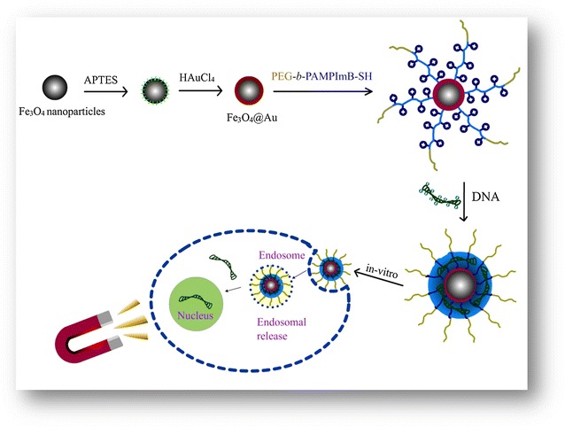
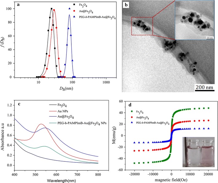
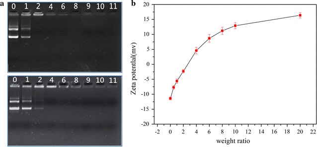
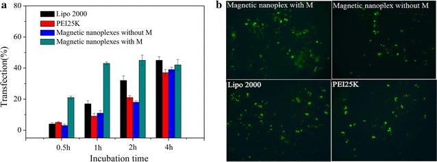
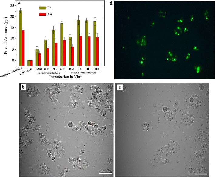
Results: Heterogeneous Au-coated Fe3O4 (Fe3O4@Au) NPs capped by polyethylene glycol-b-poly1-(3-aminopropyl)-3-(2-methacryloyloxy propylimidazolium bromine) (PEG-b-PAMPImB-Fe3O4@Au) were prepared for DNA loading and magnetofection assays. The Au outer shell of the NPs is an effective platform for maintaining the superparamagnetism of Fe3O4 and for PEG-b-PAMPImB binding via Au-S covalent bonds. By forming an electrostatic complex with DNA at the inner PAMPImB shell, the magnetic nanoplexes offer steric protection from the outer corona PEG, thereby promoting high colloidal stability. Transfection efficiency assays in human esophageal cancer cells (EC109) show that the nanoplexes have high transfection efficiency at a short incubation time in the presence of an external magnetic field, due to increased cellular internalization via magnetic acceleration. Finally, after transfection with the magnetic nanoplexes EC109 cells acquire magnetic properties, thus allowing for selective separation of transfected cells.

Conclusion: Precisely engineered architectures based on neutral-cationic block copolymer-conjugated heterogeneous NPs provide a valuable strategy for improving the applicability and efficacy of synthesized vectors.

Keywords: Block copolymer; Colloidal stability; Gene vector; Heterogeneous nanoparticles; Magnetofection.


Figures

Scheme 1
Scheme 1
Schematic representation of the magnetic vector and magnetofection process
Fig. 1
Fig. 1
Hydrodynamic diameter distributions of Fe3O4 NPs, Fe3O4@Au NPs and PEG-b-PAMPImB-Fe3O4@Au NPs (a); TEM image of PEG-b-PAMPImB-Fe3O4@Au NPs (b); the inserted image is a magnification of the NPs in b; UV–vis spectra of Fe3O4 NPs, Au NPs, Fe3O4@Au NPs and PEG-b-PAMPImB-Fe3O4@Au NPs (c); magnetization measurements as a function of applied field for Fe3O4 NPs, Fe3O4@Au NPs and PEG-b-PAMPImB-Fe3O4@Au NPs (d). The inserted image in d is a photograph of a solution of PEG-b-PAMPImB-Fe3O4@Au NPs before (right bottle) and after (left bottle) applying a NdFeB magnet during 30 min
Fig. 2
Fig. 2
Gel migration assay for PEG-b-PAMPImB (up) and PEG-b-PAMPImB-Fe3O4@Au (down) complex with DNA at various weight ratios (a), where the number on each lane represents the ratio of PEG-b-PAMPImB and PEG-b-PAMPImB-Fe3O4@Au to DNA in complexes; zeta potentials of PEG-b-PAMPImB-Fe3O4@Au/DNA complexes in pure water at various weight ratios (b). Values represent mean (±SD [n = 3])
Fig. 3
Fig. 3
a Cytotoxicity of PEI25k, PEG-b-PAMPImB and PEG-b-PAMPImB-Fe3O4@Au in EC109 cells at various concentrations; b cytotoxicity of PEG-b-PAMPImB-Fe3O4@Au/DNA in HepG2, HeLa and EC109 cells at different weight ratios. Values represent mean (SD [n = 3])
Fig. 4
Fig. 4
Transfection efficiency (% of cells transfected) of EC109 cells treated with pEGFP-C1 and magnetic nanoplexes with or without application of a magnetic field, PEI (25 kD) and Lipo2000 at various incubation times (a); fluorescence microscope images of the EC109 cells in a at 1 h of incubation (b). Values represent mean (SD [n = 3])
Fig. 5
Fig. 5
The Fe and Au content in transfected cells at various incubation times (a); confocal microscopic images of EC109 cells transfected with magnetic nanoplex with (b) and without the application of a magnetic filed (c) at 1 h incubation time (scale bar is 50 μm); fluorescence microscope image of EC109 transfected cells after magnetic collection and growth overnight (d). Values represent mean (SD [n = 3])

References

    1. Ji W, Sun B, Su C. Targeting MicroRNAs in cancer gene therapy. Genes. 2017;8:21. doi: 10.3390/genes8010021. - DOI - PMC - PubMed
    1. Hendricks WPD, Yang J, Sur S, Zhou S. Formulating the magic bullet: barriers to clinical translation of nanoparticle cancer gene therapy. Nanomedicine. 2014;9:1121–1124. doi: 10.2217/nnm.14.63. - DOI - PubMed
    1. Grigsby CL, Ho Y-P, Leong KW. Understanding nonviral nucleic acid delivery with quantum dot-FRET nanosensors. Nanomedicine. 2012;7:565–577. doi: 10.2217/nnm.12.28. - DOI - PMC - PubMed
    1. Cavallaro G, Licciardi M, Scirè S, Giammona G. Microwave-assisted synthesis of PHEA-oligoamine copolymers as potential gene delivery systems. Nanomedicine. 2009;4:291–303. doi: 10.2217/nnm.09.7. - DOI - PubMed
    1. Nayerossadat N, Maedeh T, Ali PA. Viral and nonviral delivery systems for gene delivery. Adv Biomed Res. 2012;1:27. doi: 10.4103/2277-9175.98152. - DOI - PMC - PubMed