Review
. 2017 Sep:69:140-150.
doi: 10.1016/j.semcdb.2017.07.023. Epub 2017 Jul 22.

Structural origins of clustered protocadherin-mediated neuronal barcoding

Affiliations
Review

Structural origins of clustered protocadherin-mediated neuronal barcoding

Rotem Rubinstein et al. Semin Cell Dev Biol. 2017 Sep.

Abstract

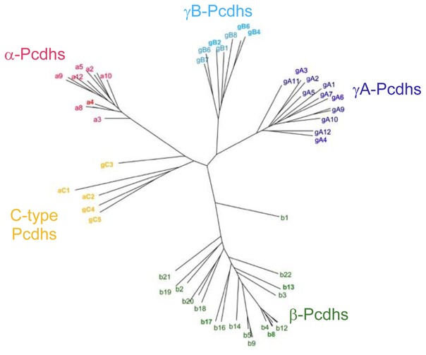
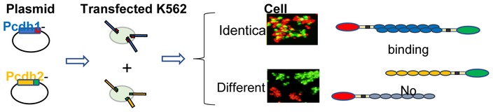
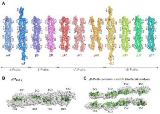
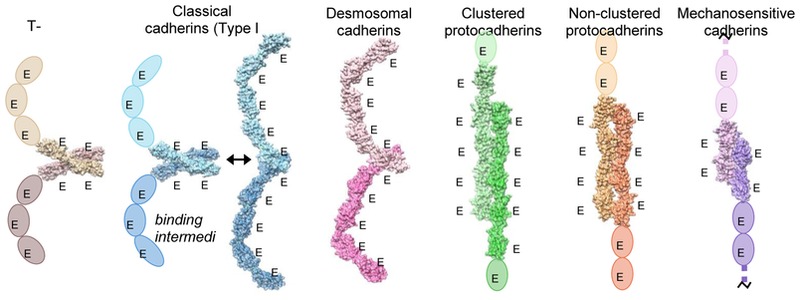
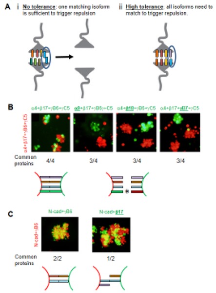
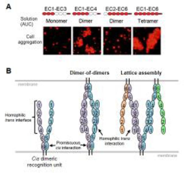
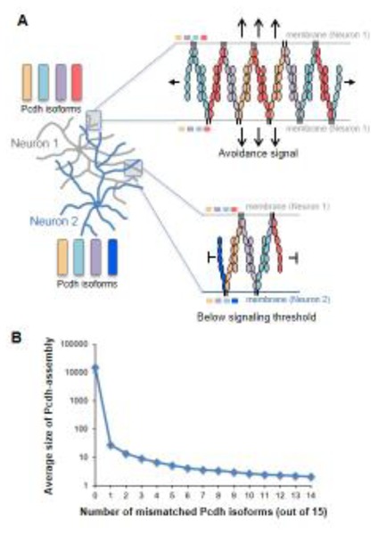
Clustered protocadherins mediate neuronal self-recognition and non-self discrimination-neuronal "barcoding"-which underpin neuronal self-avoidance in vertebrate neurons. Recent structural, biophysical, computational, and cell-based studies on protocadherin structure and function have led to a compelling molecular model for the barcoding mechanism. Protocadherin isoforms assemble into promiscuous cis-dimeric recognition units and mediate cell-cell recognition through homophilic trans-interactions. Each recognition unit is composed of two arms extending from the membrane proximal EC6 domains. A cis-dimeric recognition unit with each arm coding adhesive trans homophilic specificity can generate a zipper-like assembly that in turn suggests a chain termination mechanism for self-vs-non-self-discrimination among vertebrate neurons.

Keywords: Cell-cell recognition; Clustered protocadherins; Crystal structure; Neuronal self-avoidance; Protein interaction specificity.


Figures

Figure 1
Figure 1
Phylogenetic tree of mouse Pcdh isoforms based on the sequences of their EC1–EC4 domains which mediate the homophilic trans-interactions. The a, b, gA, and gB isoforms are grouped into four separate clusters. Although part of the b-cluster, the b1 isoform is significantly divergent from other members of the b-cluster. The two aC and the three gC isoforms are divergent from all other isoforms, including the other members of the a and g clusters
Figure 2
Figure 2
Schematic diagram of the cell aggregation assays used to assess the trans-binding properties of Pcdh isoforms. The binding properties of two exemplary isoforms are summarized. Cells transfected with Pcdh isoforms tagged with either red or green fluorescent proteins are mixed. Aggregates containing both red and green cells form when both cell populations are expressing identical isoforms. Separate red and green aggregates form when the two cell populations are expressing different isoforms, demonstrating Pcdh trans interactions are preferentially homophilic.
Figure 3
Figure 3
A) Trans-dimer structures of representative isoforms from the a, b, gA and gB clusters. The structures are shown in ribbon depiction with transparent molecular surfaces. Bound calcium ions are shown as green spheres. Glycans are shown as red, white, and blue spheres. B) Surface view of the gB7 trans-dimer. C) Open book depiction of the gB7 dimer revealing the interacting faces. Interfacial residues are colored grey if they are constant among all gB isoforms or colored green if they vary among gB isoforms. The EC1/EC4 interaction was absent in the gA8 crystal structure and one of two dimers observed in the gA1 crystal structure. While this suggests some flexibility in Pcdh structure, the lack of the EC1/EC4 interactions is likely due to crystallization artifacts since mutagenesis studies show that all four domains are required for dimerization and cell-cell recognition for these isoforms
Figure 4
Figure 4
Trans-binding interactions of cadherin superfamily proteins. Representative crystal structures of the trans-binding mode of each cadherin family for which there is structural information are shown. Crystal structures are shown as molecular surfaces, which each protomer engaged in the dimer interaction colored in a light or dark shade. Extracellular domains that are not present in the crystal structure are shown as ovals. T-cadherin PDB:3K5S [51]; Type I classical cadherin X-dimer encounter complex, E-cadherin W2A mutant PDB:3LNH [52]; Type I classical cadherin strand-swap interface, C-cadherin PDB:1L3W; Desmosomal cadherin desmocolin1 PDB:5IRY [46]; Clustered protocadherin a7 PDB:5DZV [47]; Non-clustered d-protocadherin, protocadherin 19 PDB:5IU9 [54]; Tip-link proteins, cadherin-23 (dark purple) and protocadherin-15 (light purple), PDB:4APX [53].
Figure 5
Figure 5
Tolerance and Interference. A) Schematic diagrams illustrating two extreme ‘tolerance to common isoforms’ levels. i) With no tolerance the recognition of one isoform in common between two interacting neurons is sufficient to mediate Pcdh recognition and to trigger repulsion even when all other isoforms are different. ii) With high tolerance one Pcdh mismatch between two interacting neurons is sufficient to interfere with Pcdhs recognition and ultimately repulsion even if all other Pcdhs isoforms are the same. B) Aggregation assays with K562 cells transfected with four Pcdh isoforms labeled in green or red. The underlined isoform in green indicates a mismatched isoform between the red and the green cells, which, as can be seen, results in the red and green cells forming separate aggregates. Experiments with two to five transfected isoforms gave similar results [36]. C) Control assays showing that N-cadherin does not “interfere” with Pcdh adhesion.
Figure 6
Figure 6
A) Analytical ultracentrifugation (AUC) and cell aggregation results for Pcdh gB6 domain deletion constructs. Domains that are present are shown in red. B) Alternative models for Pcdh trans interactions based on a cis dimeric recognition units (left). In the discrete tetramer model (center), a “two-armed” trans dimer forms between identical cis dimers. In the lattice model, cis-dimeric recognition units interact to form a zipper-like structure.
Figure 7
Figure 7
Chain termination model for neuronal recognition based on a Pcdh zipper-assembly. A) Top – For apposed neurites originating from the same neuron shown with four identical isoforms, Pcdh zipper assembly is limited only by Pcdh copy number. Bottom – For apposed neurites from different neurons with single isoform mismatch chain termination occurs quickly, through incorporation into the assembly of the mismatched isoforms. B) Monte-Carlo calculations demonstrate a striking dependence of Pcdh assembly size on the number of mismatched isoforms between interacting cells. Calculations assume 1000 copies per isoform. Note the step-function shape of the curve.

References

    1. Azevedo FA, Carvalho LR, Grinberg LT, Farfel JM, Ferretti RE, Leite RE, et al. Equal numbers of neuronal and nonneuronal cells make the human brain an isometrically scaled-up primate brain. J Comp Neurol. 2009;513:532–41. - PubMed
    1. Lefebvre JL, Kostadinov D, Chen WV, Maniatis T, Sanes JR. Protocadherins mediate dendritic self-avoidance in the mammalian nervous system. Nature. 2012;488:517–21. - PMC - PubMed
    1. Kostadinov D, Sanes JR. Protocadherin-dependent dendritic self-avoidance regulates neural connectivity and circuit function. Elife. 2015:4. - PMC - PubMed
    1. Mountoufaris G, Chen WV, Hirabayashi Y, O’Keeffe S, Chevee M, Nwakeze CL, et al. Multicluster Pcdh diversity is required for mouse olfactory neural circuit assembly. Science. 2017;356:411–4. - PMC - PubMed
    1. Chen WV, Alvarez FJ, Lefebvre JL, Friedman B, Nwakeze C, Geiman E, et al. Functional Significance of Isoform Diversification in the Protocadherin Gamma Gene Cluster. Neuron. 2012;75:402–9. - PMC - PubMed

Publication types

LinkOut - more resources