

The effects of Suramin on Ca2+ activated force and sarcoplasmic reticulum Ca2+ release in skinned fast-twitch skeletal muscle fibers of the rat
- PMID: 28743820
- PMCID: PMC5532480
- DOI: 10.14814/phy2.13333
The effects of Suramin on Ca2+ activated force and sarcoplasmic reticulum Ca2+ release in skinned fast-twitch skeletal muscle fibers of the rat
Abstract
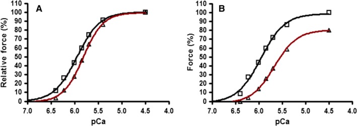
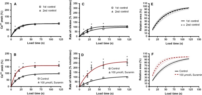
Suramin has long been used in the treatment of various human diseases. Intravenous infusions of Suramin are commonly administered to patients over extended periods of time but there are a number of significant contraindications with peripheral muscle weakness being one of the most frequently reported. Previous work has shown that even after a single infusion (300 mg kg-1) Suramin remains in skeletal muscle in effective concentrations (11.6 μg mL-1; 84 days) for prolonged periods. These observations provide a strong rationale for investigation of the specific effects of Suramin on skeletal muscle function. Single mechanically skinned fibers were directly exposed to Suramin (10, 100 or 500 μmol L-1) for defined durations (2-10 min) in controlled physiological solutions that mimic the intracellular ionic environment of a fiber. Suramin treatment (10-500 μmol L-1) directly affected the contractile apparatus in a dose-dependent manner causing a decrease in Ca2+-sensitivity (pCa50 = -log (Ca2+) concentration, where 50% of maximum Ca2+- activated force is produced) by 0.14 to 0.42 pCa units and reduction in maximum Ca2+-activated force by 14 to 62%. Suramin treatment (100 μmol L-1 for 10 min and 500 μmol L-1 for 2 min) also caused development of a Ca2+-independent force corresponding to 2.89 ± 4.33 and 16.77 ± 7.50% of pretreatment maximum Ca2+-activated force, respectively. Suramin treatment (100 μmol L-1, 2 min) also increased the rate of sarcoplasmic reticulum (SR) Ca2+ release without significant changes in SR Ca2+ uptake. We report new functional effects for Suramin related to alterations in both the contractile apparatus and SR Ca2+-handling of skeletal muscle that may contribute to the peripheral muscle weakness noted in human pharmacological treatments.
Keywords: EC‐coupling; mechanically skinned; skeletal muscle; suramin.
© 2017 The Authors. Physiological Reports published by Wiley Periodicals, Inc. on behalf of The Physiological Society and the American Physiological Society.
Figures


Similar articles
-
Effects of elevated physiological temperatures on sarcoplasmic reticulum function in mechanically skinned muscle fibers of the rat.Am J Physiol Cell Physiol. 2007 Jul;293(1):C133-41. doi: 10.1152/ajpcell.00052.2007. Epub 2007 Mar 7. Am J Physiol Cell Physiol. 2007. PMID: 17344316
-
Contractile properties and sarcoplasmic reticulum calcium content in type I and type II skeletal muscle fibres in active aged humans.J Physiol. 2015 Jun 1;593(11):2499-514. doi: 10.1113/JP270179. Epub 2015 Apr 17. J Physiol. 2015. PMID: 25809942 Free PMC article.
-
The effect of 2,5-di-(tert-butyl)-1,4-hydroquinone on force responses and the contractile apparatus in mechanically skinned muscle fibres of the rat and toad.J Muscle Res Cell Motil. 1996 Feb;17(1):55-67. doi: 10.1007/BF00140324. J Muscle Res Cell Motil. 1996. PMID: 8740432
-
Measurement of force and calcium release using mechanically skinned fibers from mammalian skeletal muscle.J Appl Physiol (1985). 2018 Oct 1;125(4):1105-1127. doi: 10.1152/japplphysiol.00445.2018. Epub 2018 Jul 19. J Appl Physiol (1985). 2018. PMID: 30024333 Review.
-
A study of the mechanisms of excitation-contraction coupling in frog skeletal muscle based on measurements of [Ca2+] transients inside the sarcoplasmic reticulum.J Muscle Res Cell Motil. 2018 Apr;39(1-2):41-60. doi: 10.1007/s10974-018-9497-9. Epub 2018 Aug 24. J Muscle Res Cell Motil. 2018. PMID: 30143958 Review.
Cited by
-
The effect of intrauterine growth restriction on Ca2+ -activated force and contractile protein expression in the mesenteric artery of adult (6-month-old) male and female Wistar-Kyoto rats.Physiol Rep. 2018 Dec;6(24):e13954. doi: 10.14814/phy2.13954. Physiol Rep. 2018. PMID: 30592188 Free PMC article.
References
-
- Allhouse, L. D. , Potter J. D., and Ashley C. C.. 1999. A novel method of extraction of TnC from skeletal muscle myofibrils. Pflugers Arch. 437:695–701. - PubMed
-
- Eisenberger, M. A. , Sinibaldi V., and Reyno L.. 1995. Suramin. Cancer Pract. 3:187–189. - PubMed
-
- Emmick, J. T. , Kwon S., Bidasee K. R., Besch K. T., and Besch H. R. Jr. 1994. Dual effect of suramin on calcium fluxes across sarcoplasmic reticulum vesicle membranes. J. Pharmcol. Exp. Ther. 269:717–724. - PubMed
-
- Endo, M. , and Iino M.. 1980. Specific Perforation of muscle cell membranes with preserved SR functions by saponin treatment. J. Muscle Res. Cell Motil. 1:89–100. - PubMed
MeSH terms
Substances
LinkOut - more resources
Full Text Sources
Other Literature Sources
Research Materials
Miscellaneous
