. 2017 Jul 25;7(1):6386.
doi: 10.1038/s41598-017-06281-1.

Transcriptome Analysis of Canine Cutaneous Melanoma and Melanocytoma Reveals a Modulation of Genes Regulating Extracellular Matrix Metabolism and Cell Cycle

Affiliations

Transcriptome Analysis of Canine Cutaneous Melanoma and Melanocytoma Reveals a Modulation of Genes Regulating Extracellular Matrix Metabolism and Cell Cycle

Chiara Brachelente et al. Sci Rep. .

Abstract

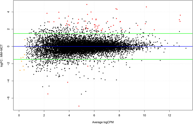
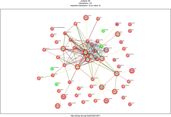
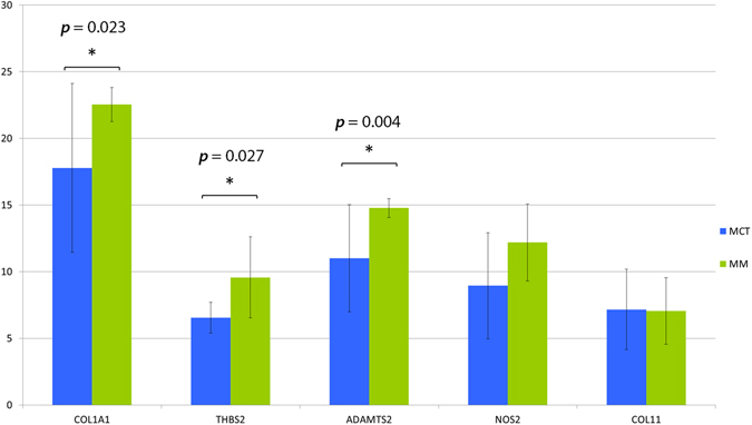
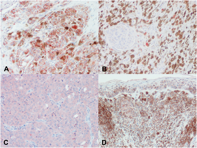
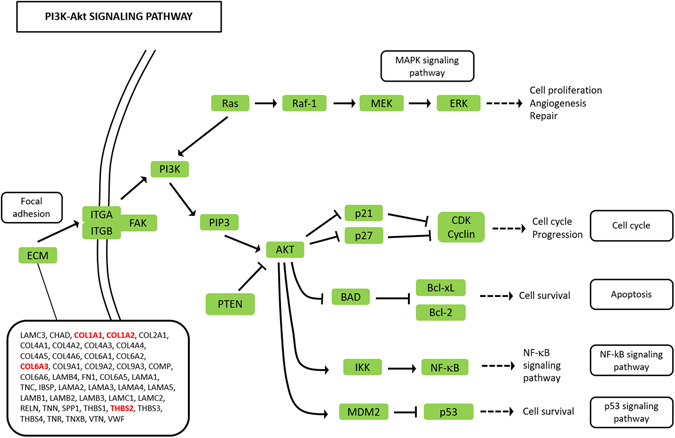
Interactions between tumor cells and tumor microenvironment are considered critical in carcinogenesis, tumor invasion and metastasis. To examine transcriptome changes and to explore the relationship with tumor microenvironment in canine cutaneous melanocytoma and melanoma, we extracted RNA from formalin-fixed, paraffin-embedded (FFPE) specimens and analyzed them by means of RNA-seq for transcriptional analysis. Melanocytoma and melanoma samples were compared to detect differential gene expressions and significant enriched pathways were explored to reveal functional relations between differentially expressed genes. The study demonstrated a differential expression of 60 genes in melanomas compared to melanocytomas. The differentially expressed genes cluster in the extracellular matrix-receptor interaction, protein digestion and absorption, focal adhesion and PI3K-Akt (phosphoinositide 3-kinase/protein kinase B) signaling pathways. Genes encoding for several collagen proteins were more commonly differentially expressed. Results of the RNA-seq were validated by qRT-PCR and protein expression of some target molecules was investigated by means of immunohistochemistry. We hypothesize that the developing melanoma actively promotes collagen metabolism and extracellular matrix remodeling as well as enhancing cell proliferation and survival contributing to disease progression and metastasis. In this study, we also detected unidentified genes in human melanoma expression studies and uncover new candidate drug targets for further testing in canine melanoma.


Conflict of interest statement

The authors declare that they have no competing interests.

Figures

Figure 1
Figure 1
MA plot showing the relationship between average concentration (logCPM) and fold-change (logFC) across the genes. Each gene is represented by a black dot. Significant differentially expressed genes are colored in red. The orange dots represent genes in which the counts were zero in all samples of one of the groups. The green lines represent logFC +/− 1.5 threshold.
Figure 2
Figure 2
Network of the differentially expressed genes produced by String. Circles are proteins while lines are interactions. Known interactions are represented in purple (experimentally determined) and azure (from curated databases). Red, green and blue lines pertain predicted interactions (respectively gene neighborhood, fusions and co-occurrence) while yellow, indigo and black are interactions found from other sources (text mining, co-expression and protein homology). The red halo around gene represents up-regulation while the green one down-regulation.
Figure 3
Figure 3
Bar charts representing relative expression values of COL1A1, THBS2, ADAMTS2, NOS2 and COL11 genes in melanocytomas (blue) and melanoma (green) on a base 2 logarithmic scale. Whiskers define a 95% CI (confidence interval). Statistical significance is evidenced with asterisks and relative p-value.
Figure 4
Figure 4
Immunohistochemical expression of COL1 and THBS2 in melanocytic tumors. (A) Cytoplasmic positivity for COL1 in numerous neoplastic cells and fewer stromal cells in a melanoma; (B) occasional COL1 positive cells in melanocytoma; (C) diffuse cytoplasmic stain for THBS2 in an amelanotic melanoma; (D) scattered and mild THBS2 positivity in melanocytoma cells.
Figure 5
Figure 5
The extended Kegg Pathway for PI3K-Akt signaling. Genes activating the ECM-receptor interaction pathway and upstream of the PI3K-Akt molecule are depicted in the rectangle. The genes that were differentially expressed in this study are bold face and red colored.

Similar articles

Cited by

References

    1. Balch CM, et al. Final version of 2009 AJCC melanoma staging and classification. J. Clin. Oncol. 2009;27:6199–206. doi: 10.1200/JCO.2009.23.4799. - DOI - PMC - PubMed
    1. Hoek K, et al. Expression profiling reveals novel pathways in the transformation of melanocytes to melanomas expression profiling reveals novel pathways in the transformation of melanocytes to melanomas. Cancer Res. 2004;165:5270–5282. doi: 10.1158/0008-5472.CAN-04-0731. - DOI - PubMed
    1. Smith AP, Hoek K, Becker D. Whole-genome expression profiling of the melanoma progression pathway reveals marked molecular differences between nevi/melanoma in situ and advanced-stage melanomas. Cancer Biol. Ther. 2005;4:1018–1029. doi: 10.4161/cbt.4.9.2165. - DOI - PubMed
    1. Stuelten CH, et al. Breast cancer cells induce stromal fibroblasts to express MMP-9 via secretion of TNF-alpha and TGF-beta. J. Cell Sci. 2005;118:2143–53. doi: 10.1242/jcs.02334. - DOI - PubMed
    1. Hanahan D, Weinberg RA. Hallmarks of cancer: the next generation. Cell. 2011;144:646–74. doi: 10.1016/j.cell.2011.02.013. - DOI - PubMed

Publication types

MeSH terms