. 2017 Jul 25;8(1):125.
doi: 10.1038/s41467-017-00200-8.

Long-term hepatitis B infection in a scalable hepatic co-culture system

Affiliations

Long-term hepatitis B infection in a scalable hepatic co-culture system

Benjamin Y Winer et al. Nat Commun. .

Abstract

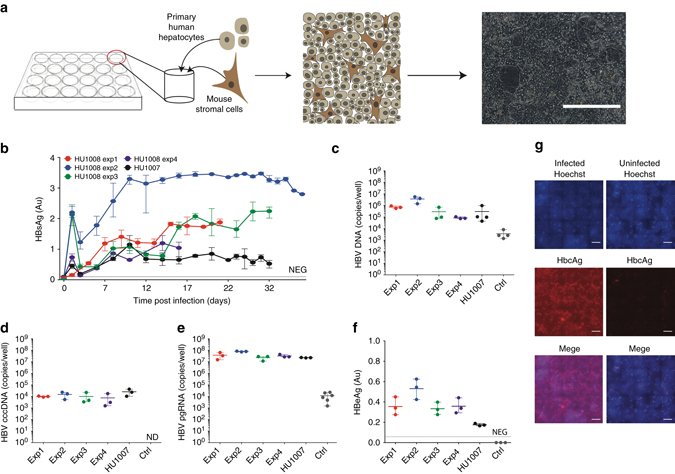
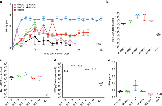
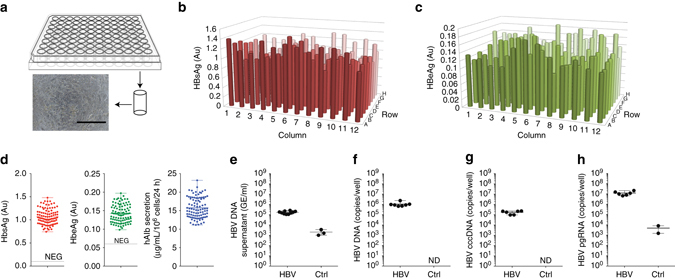
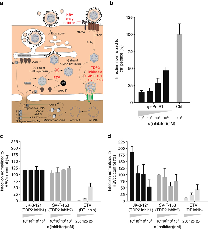
Hepatitis B virus causes chronic infections in 250 million people worldwide. Chronic hepatitis B virus carriers are at risk of developing fibrosis, cirrhosis, and hepatocellular carcinoma. A prophylactic vaccine exists and currently available antivirals can suppress but rarely cure chronic infections. The study of hepatitis B virus and development of curative antivirals are hampered by a scarcity of models that mimic infection in a physiologically relevant, cellular context. Here, we show that cell-culture and patient-derived hepatitis B virus can establish persistent infection for over 30 days in a self-assembling, primary hepatocyte co-culture system. Importantly, infection can be established without antiviral immune suppression, and susceptibility is not donor dependent. The platform is scalable to microwell formats, and we provide proof-of-concept for its use in testing entry inhibitors and antiviral compounds.The lack of models that mimic hepatitis B virus (HBV) infection in a physiologically relevant context has hampered drug development. Here, Winer et al. establish a self-assembling, primary hepatocyte co-culture system that can be infected with patient-derived HBV without further modifications.


Conflict of interest statement

E.P., Am.P., C.C., A.S., and E.N. are employees of the Hurel Corporation of which E.N. is also a stockholder. The remaining authors declare no competing financial interests.

Figures

Fig. 1
Fig. 1
Persistent HBV infection of self-assembling primary hepatocyte co-cultures. a PHH platform. HBVcc infection of mixed PHH donors performed in five separate experiments with two separate lots of hepatocytes. HBsAg concentrations determined in the supernatants b and total HBV DNA c, HBV cccDNA d, HBV pgRNA e quantified in SACC-PHH lysates at the final HBsAg timepoint. f HBeAg was quantified in the supernatants (day 16 post infection) by HBeAg ELISA. g HBcAg detection in HBV-infected (left panels) and non-infected (right panels) mixed donor SACC-PHHs by immunofluorescence microscopy; HBcAg (red), nuclear Hoechst dye (blue), all scale bars are 200 μm. For all HBVcc infections of mixed PHH donors three to five biological replicates were performed. All data are presented as means ± s.d
Fig. 2
Fig. 2
Persistent HBV infection in single donor PHH co-cultures. Assessment of HBVcc infection of single donor SACC-PHH co-cultures. Quantification of HBsAg concentrations in the supernatants a, and total HBV DNA b, cccDNA c, pgRNA d in SACC-PHH lysates. e HBeAg was quantified in the supernatants (day 16 post infection) by HBeAg ELISA. For all HBVcc infections of single PHH donors three to five biological replicates were performed. All data are presented as means ± s.d
Fig. 3
Fig. 3
Long-term persistent infection of SACC-PHHs with patient-derived HBV. Assessment of heparin column-purified HBVpat infection of mixed donor HU1008 SACC-PHHs. Quantification of HBsAg concentrations in the supernatants a, and total HBV DNA b, cccDNA c, and pgRNA d in SACC-PHH lysates. For all HBVpat infections of mixed donors four biological replicates were performed. All data are presented as means ± s.d
Fig. 4
Fig. 4
Robust HBV infection SACC-PHHs in microwell formats. a Schematic depiction and representative bright-field image of the microwell SACC-PHH system (scale bar = 400 μm). Quantification of HBsAg b and HBeAg c across the 96-well format at 10 dpi (day 20 post seeding). d Limited variation of HBsAg (left, mean 1.070 Au, std 0.167 Au, two tail t-test p-value < 0.0001, compared to 1 Au), HBeAg (middle, mean 0.142 Au, std 0.024 Au, two tail t-test p-value < 0.0001, compared to 0 Au) and hAlb (right, mean 15.75 μgml per 106 cells per 24 h, std 2.679 μgml per 106 cells per 24 h, two tail t-test p-value < 0.001, compared to 15 μgml per106 cells per 24 h) at 10 dpi. Quantification of HBV DNA in culture supernatants e at 10 dpi, and total HBV DNA f, cccDNA g and pgRNA h in cell lysates of randomly picked wells at 30 dpi. For HBV DNA and RNA quantifications six to ten replicates were performed. For HBsAg, HBeAg, and hAlb 96-biological replicates were performed. All data are presented as means ± s.d
Fig. 5
Fig. 5
Utility of SACC-PHHs for antiviral drug testing. a Schematic of HBV life cycle indicating the presumed mechanism of action of myr-PreS1 entry inhibitor, TDP2 inhibitors and ETV. b Prophylactic treatment with myr-preS1-derived peptides in SACC-PHH 96-well format (HU1007 mixed donor). Prophylactic c and therapeutic d drug dosing of SACC-PHHs (mixed donor HU1008) in 96-well format for nucleotide analog ETV and TDP2 inhibitors (JK-3-121, SV-F-153), x-axis: concentration of different drugs. y-axis: amount of HBsAg secretion normalized to that secreted by HBVcc infected untreated control cells. For HBVcc infections and drug treatments, six biological replicates were performed. All data are presented as means ± s.d

References

    1. Ni Y, et al. Hepatitis B and D viruses exploit sodium taurocholate co-transporting polypeptide for species-specific entry into hepatocytes. Gastroenterology. 2014;146:1070–1083. doi: 10.1053/j.gastro.2013.12.024. - DOI - PubMed
    1. Yan H, et al. Sodium taurocholate cotransporting polypeptide is a functional receptor for human hepatitis B and D virus. eLife. 2012;1:e00049. doi: 10.7554/eLife.00049. - DOI - PMC - PubMed
    1. Li W, Urban S. Entry of hepatitis B and hepatitis D virus into hepatocytes: basic insights and clinical implications. J. Hepatol. 2016;64:S32–S40. doi: 10.1016/j.jhep.2016.02.011. - DOI - PMC - PubMed
    1. Winer BY, Ploss A. Determinants of hepatitis B and delta virus host tropism. Curr. Opin. Virol. 2015;13:109–116. doi: 10.1016/j.coviro.2015.06.004. - DOI - PMC - PubMed
    1. Guo H, et al. Characterization of the intracellular deproteinized relaxed circular DNA of hepatitis B virus: an intermediate of covalently closed circular DNA formation. J. Virol. 2007;81:12472–12484. doi: 10.1128/JVI.01123-07. - DOI - PMC - PubMed

Publication types

MeSH terms

Substances