. 2017 Jul 25;7(1):6361.
doi: 10.1038/s41598-017-06610-4.

NADPH oxidase 4 is required for the generation of macrophage migration inhibitory factor and host defense against Toxoplasma gondii infection

Affiliations

NADPH oxidase 4 is required for the generation of macrophage migration inhibitory factor and host defense against Toxoplasma gondii infection

Ji Hye Kim et al. Sci Rep. .

Abstract

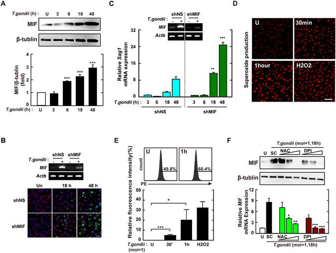
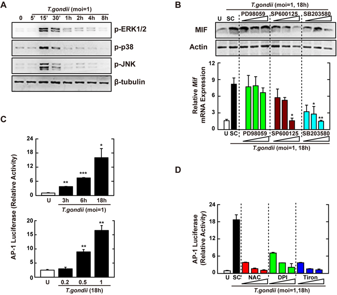
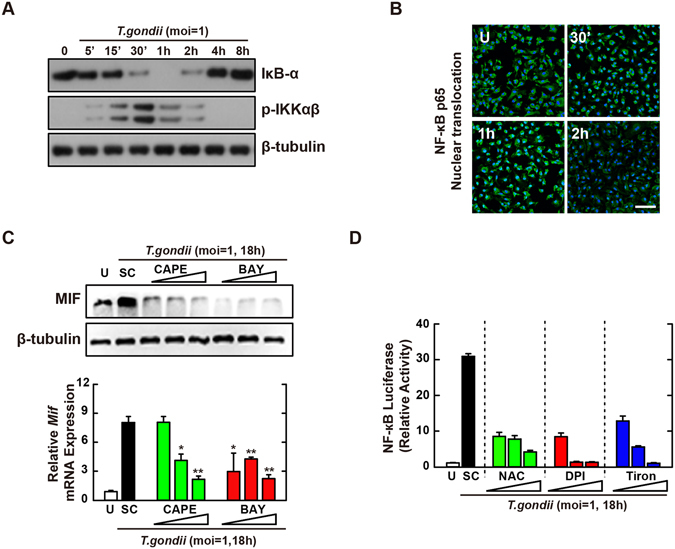
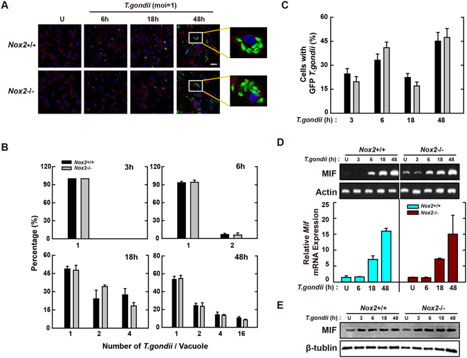
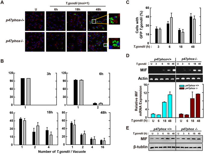
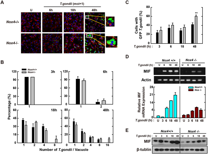
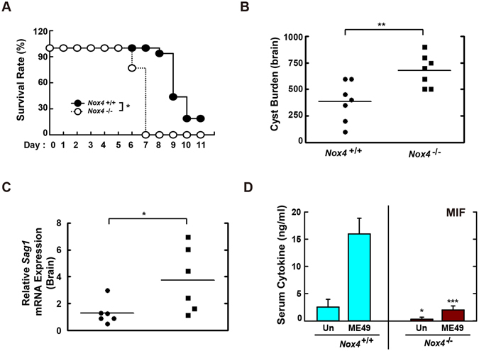
Nicotinamide adenine dinucleotide phosphate (NADPH) oxidases (Nox) are an important family of catalytic enzymes that generate reactive oxygen species (ROS), which mediate the regulation of diverse cellular functions. Although phagocyte Nox2/gp91phox is closely associated with the activation of host innate immune responses, the roles of Nox family protein during Toxoplasma gondii (T. gondii) infection have not been fully investigated. Here, we found that T. gondii-mediated ROS production was required for the upregulation of macrophage migration inhibitory factor (MIF) mRNA and protein levels via activation of mitogen-activated protein kinase and nuclear factor-κB signaling in macrophages. Interestingly, MIF knockdown led to a significant increase in the survival of intracellular T. gondii in bone marrow-derived macrophages (BMDMs). Moreover, Nox4 deficiency, but not Nox2/gp91phox and the cytosolic subunit p47phox, resulted in enhanced survival of the intracellular T. gondii RH strain and impaired expression of T. gondii-mediated MIF in BMDMs. Additionally, Nox4-deficient mice showed increased susceptibility to virulent RH strain infection and increased cyst burden in brain tissues and low levels of MIF expression following infection with the avirulent ME49 strain. Collectively, our findings indicate that Nox4-mediated ROS generation plays a central role in MIF production and resistance to T. gondii infection.


Conflict of interest statement

The authors declare that they have no competing interests.

Figures

Figure 1
Figure 1
MIF expression via intracellular ROS generation is required for the inhibition of intracellular proliferation in T. gondii-infected macrophages. (A) BMDMs were infected with T.gondii RH strain (moi = 1) for the indicated time periods and then cell lysate was collected. Immunoblot analysis was performed for protein expression of MIF or β-tubulin. Upper panel, Representative gel image. Lower panel, Densitometry. (B,C) BMDM were transduced with lentiviruses expressing shNS or shMIF at a multiplicity of infection (MOI) of 5 for 48 h with polybrene (8 μg/mL) and then infected with GFP-RH strain (for B) or T. gondii RH strain (for C) for the indicated time periods. The mRNA expression for Mif and Actb was determined using semiquantitative RT-PCR (B) Cells were fixed and stained with Texas Red®-X phalloidin for labeling F-actin (red) for cytosolic fraction, and DAPI (blue) for nuclei and then analyzed for the number of GFP-RH strain using confocal microscopy (bottom) (C) Quantitative real-time PCR analysis were assessed to determine sag1 mRNA expression in whole-cell lysates. (D,E) BMDMs were infected with T. gondii RH strain (moi = 1) for indicated times and then stained with DHE (2 μM) for 15 min. Intracellular ROS generation was measured using confocal microscopy (for D) and flow cytometery (for E). Scale bar = 50 μm. H2O2 (1 mM, 30 min) was used for positive control. (F) Immunoblot (top) or qPCR (bottom) analysis of MIF expression in BMDMs after T. gondii RH strain infection (moi = 1, 18 hr) in the presence or absence of general antioxidant (NAC; 1, 2, or 5 mM) or Nox inhibitor (DPI; 1, 5, or 10 μM). Data are representative of three independent experiments and are presented as means ± SD. *P < 0.05, **P < 0.01, ***P < 0.001, two-tailed Student’s t-test.
Figure 2
Figure 2
T. gondii-induced MIF expression is mediated through the activation of JNK and p38 MAPK pathway, followed by the AP-1 transcriptional activation. (A) BMDMs were infected with T. gondii RH strain (moi = 1) for the indicated time periods. Cells were harvested and subjected to western blotting analysis for phosphorylated ERK, p38, or JNK. β-tubulin served as a loading control. (B) BMDMs were pretreated with PD98059 (5, 10, 20 μM), SP600125 (5, 20, 30 μM) or SB203580 (1, 5, 10 μM) for 45 min, followed by infection with T. gondii RH strain (moi = 1) for 18 h. Immunoblot (top) or qPCR (bottom) analysis was performed to determine protein and mRNA expression MIF, respectively. (C) Raw 264.7 cells were transfected with plasmids carrying AP-1 luciferase reporter constructs before T. gondii RH strain (moi = 1, top; indicated moi, bottom) for various time periods (top) or 18 h. Luciferase assays were performed based on normalization to the β-galactosidase activity. (D) Effects on AP-1 transcriptional activity in the presence or absence of general antioxidant (NAC; 1, 2, or 5 mM), Nox inhibitor (DPI; 1, 5, or 10 μM) or superoxide scavenger (Tiron; 5, 10, or 20 mM). The experimental conditions were as outlined in Fig. 2C. Data are representative of three independent experiments and are presented as means ± SD. *P < 0.05, **P < 0.01, ***P < 0.001, two-tailed Student’s t-test.
Figure 3
Figure 3
NF-κB signaling plays an essential role in T. gondii-induced MIF expression by BMDMs. (A,B) BMDMs were infected with T. gondii RH strain (moi = 1) for the indicated time periods. (A) Immunoblot analysis was performed to determine protein expression of total IκB-α and phosphorylated IKKα/β. β-tubulin served as a loading control. (B) Immunofluorescence analyses of NF-κB p65 nuclear translocation. Cells were fixed and stained with anti-NF-κB p65 (green); nuclei were stained with DNA-intercalating dye DAPI (blue). Scale bar = 50 μm. (C) BMDMs were pretreated with CAPE (1, 5 or 10 µM, 2 h) and BAY (0.1, 1, 3 μM, 45 min) and then infected with T. gondii RH strain (moi = 1) for 18 h. Immunoblot (top) or qPCR (bottom) analysis was performed to determine protein and mRNA expression MIF, respectively. (D) Raw 264.7 cells were transfected with plasmids carrying NF-κB luciferase reporter constructs. Cells were pretreated with general antioxidant (NAC; 1, 2, or 5 mM), Nox inhibitor (DPI; 1, 5, or 10 μM) or superoxide scavenger (Tiron; 5, 10, or 20 mM) and then infected with T. gondii RH strain (moi = 1) for 18 h. Luciferase activity were measured and normalized to β-galactosidase activity. Data are representative of three independent experiments and are presented as means ± SD. *P < 0.05, **P < 0.01, ***P < 0.001, two-tailed Student’s t-test.
Figure 4
Figure 4
Macrophage-derived Nox2/gp91phox is not associated with induction of antiparasitic defense during T. gondii infection. (AC) BMDMs from Nox2 +/+ and Nox2 −/− mice were infected with GFP-RH strain (moi = 1) for the indicated time periods. (A) Cells were fixed and stained with Texas Red®-X phalloidin for labeling F-actin (red) for cytosolic fraction, and DAPI (blue) for nuclei and then analyzed for the number of GFP-RH strain using confocal microscopy (B,C) The number of GFP-RH strain per vacuole (for B) or of GFP-RH strain-infected cell (for C) were analyzed. Scale bar = 25 μm. Data are representative of five independent experiments (D,E) BMDMs from Nox2 +/+ and Nox2 −/− mice were infected with T. gondii RH strain (moi = 1) for the indicated time periods. (D) The mRNA expression for Mif and actb was determined using semiquantitative RT-PCR (top) or qPCR (bottom) analysis. (E) Immunoblot analysis was performed to determine protein expression of MIF and β-tubulin. Data are representative of three independent experiments and are presented as means ± SD.
Figure 5
Figure 5
Macrophage-derived p47phox is not involved in antiparasitic defense during T. gondii infection. (AC) BMDMs from p47phox +/+ and p47phox −/− mice were infected with GFP-RH strain (moi = 1) for the indicated time periods. (A) Cells were fixed and stained with Texas Red®-X phalloidin for labeling F-actin (red) for cytosolic fraction, and DAPI (blue) for nuclei and then analyzed for the number of GFP-RH strain using confocal microscopy (B,C) The number of GFP-RH strain per vacuole (for B) or of GFP-RH strain-infected cell (for C) were analyzed. Scale bar = 25 μm. Data are representative of five independent experiments (D,E) BMDMs from p47phox +/+ and p47phox −/− mice were infected with T. gondii RH strain (moi = 1) for the indicated time periods. (D) The mRNA expression for Mif and actb was determined using semiquantitative RT-PCR (top) or qPCR (bottom) analysis. (E) Immunoblot analysis was performed to determine protein expression of MIF and β-tubulin. Data are representative of three independent experiments and are presented as means ± SD.
Figure 6
Figure 6
Nox4 is essential for the activation of protective immunity in T. gondii-infected macrophages. (AC) BMDMs from Nox4 +/+ and Nox4 −/− mice were infected with GFP-RH strain (moi = 1) for the indicated time periods. (A) Cells were fixed and stained with Texas Red®-X phalloidin for labeling F-actin (red) for cytosolic fraction, and DAPI (blue) for nuclei and then analyzed for the number of GFP-RH strain using confocal microscopy (B,C) The number of GFP-RH strain per vacuole (for B) or of GFP-RH strain-infected cell (for C) were analyzed. Scale bar = 25 μm. Data are representative of five independent experiments (D,E) BMDMs from Nox4 +/+ and Nox4 −/− mice were infected with T. gondii RH strain (moi = 1) for the indicated time periods. (D) The mRNA expression for Mif and actb was determined using semiquantitative RT-PCR (top) or qPCR (bottom) analysis. (E) Immunoblot analysis was performed to determine protein expression of MIF and β-tubulin. Data are representative of three independent experiments and are presented as means ± SD. *P < 0.05, **P < 0.01, ***P < 0.001, two-tailed Student’s t-test.
Figure 7
Figure 7
Nox4 is required for the host resistance against both RH and ME49 strain of T. gondii. (A) Survival of Nox4 +/+ and Nox4 −/− mice (n = 13 per genotype) infected with 200 tachyzoites of T. gondii RH strain (i.p. injection). (BD) Nox4 +/+ and Nox4 −/− mice (n = 5 per genotype) were infected with 40 cysts of T. gondii ME49 strain (i.p. injection) for 20 days. (B) Number of cysts in brain of Nox4 +/+ and Nox4 −/− mice were counted using a microscope. (C) The mRNA expression for T. gondii-specific gene Sag1 in brain of Nox4 +/+ and Nox4 −/− mice was evaluated by qPCR analysis. (D) Serum MIF levels from Nox4 +/+ and Nox4 −/− mice were assessed by ELISA analysis. *P < 0.05, **P < 0.01, ***P < 0.001, compared with Nox4 +/+ mice infected with T. gondii (log-rank test (for A) or two-tailed Student’s t-test (for BD).

References

    1. Dubremetz JF. Host cell invasion by Toxoplasma gondii. Trends Microbiol. 1998;6:27–30. doi: 10.1016/S0966-842X(97)01165-7. - DOI - PubMed
    1. Elmore SA, et al. Toxoplasma gondii: epidemiology, feline clinical aspects, and prevention. Trends Parasitol. 2010;26:190–6. doi: 10.1016/j.pt.2010.01.009. - DOI - PubMed
    1. Yarovinsky F. Innate immunity to Toxoplasma gondii infection. Nat Rev Immunol. 2014;14:109–21. doi: 10.1038/nri3598. - DOI - PubMed
    1. Denkers EY. Toll-like receptor initiated host defense against Toxoplasma gondii. J Biomed Biotechnol. 2010;2010:737125. doi: 10.1155/2010/737125. - DOI - PMC - PubMed
    1. Carruthers V, Boothroyd JC. Pulling together: an integrated model of Toxoplasma cell invasion. Curr Opin Microbiol. 2007;10:83–9. doi: 10.1016/j.mib.2006.06.017. - DOI - PubMed

Publication types

MeSH terms