. 2017 Jul 25;7(1):6470.
doi: 10.1038/s41598-017-06990-7.

Permeability Barrier and Microstructure of Skin Lipid Membrane Models of Impaired Glucosylceramide Processing

Affiliations

Permeability Barrier and Microstructure of Skin Lipid Membrane Models of Impaired Glucosylceramide Processing

Michaela Sochorová et al. Sci Rep. .

Abstract

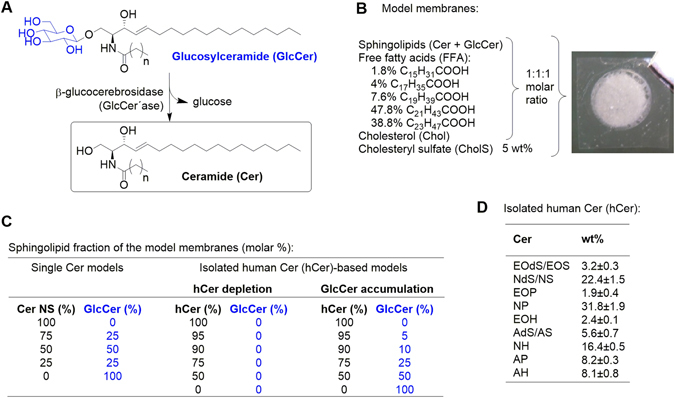
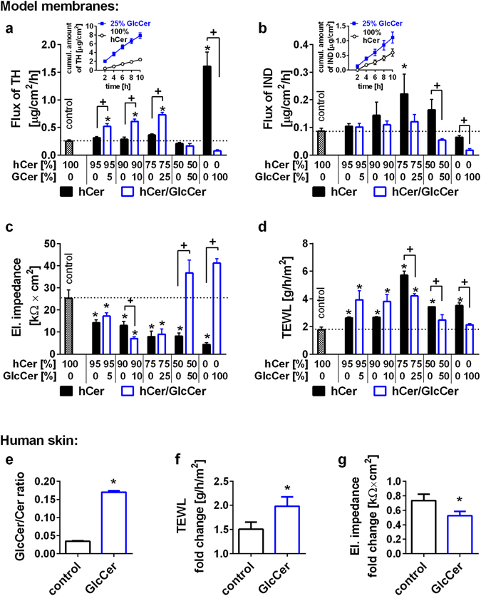
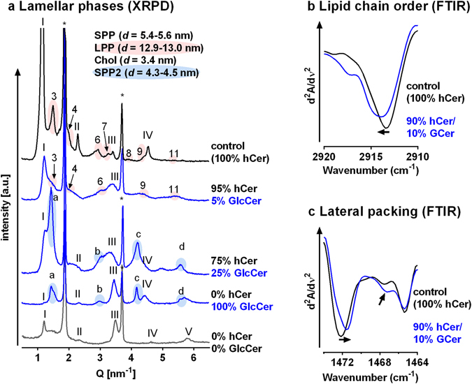
Ceramide (Cer) release from glucosylceramides (GlcCer) is critical for the formation of the skin permeability barrier. Changes in β-glucocerebrosidase (GlcCer'ase) activity lead to diminished Cer, GlcCer accumulation and structural defects in SC lipid lamellae; however, the molecular basis for this impairment is not clear. We investigated impaired GlcCer-to-Cer processing in human Cer membranes to determine the physicochemical properties responsible for the barrier defects. Minor impairment (5-25%) of the Cer generation from GlcCer decreased the permeability of the model membrane to four markers and altered the membrane microstructure (studied by X-ray powder diffraction and infrared spectroscopy), in agreement with the effects of topical GlcCer in human skin. At these concentrations, the accumulation of GlcCer was a stronger contributor to this disturbance than the lack of human Cer. However, replacement of 50-100% human Cer by GlcCer led to the formation of a new lamellar phase and the maintenance of a rather good barrier to the four studied permeability markers. These findings suggest that the major cause of the impaired water permeability barrier in complete GlcCer'ase deficiency is not the accumulation of free GlcCer but other factors, possibly the retention of GlcCer bound in the corneocyte lipid envelope.


Conflict of interest statement

The authors declare that they have no competing interests.

Figures

Figure 1
Figure 1
Scheme of the Conversion of Glucosylceramide (GlcCer) to Ceramide (Cer) by β-Glucocerebrosidase (GlcCer’ase) (A) and the Composition of the Model Membranes (BD). The model membranes were constructed from sphingolipids (Cer and/or GlcCer, Chol and a mixture of FFA in an equimolar ratio with 5 wt% CholS (B,C). For single Cer membranes, N-tetracosanoyl D-erythro-sphingosine (CerNS) was used, whereas the complex model system was constructed from isolated human SC Cer (hCer). The composition of hCer is specified in panel D. To simulate deficient GlcCer-to-Cer processing, the Cer fraction was gradually diminished with or without GlcCer as a replacement (C).
Figure 2
Figure 2
Permeabilities of the hCer Membrane Models (ad) and Effects of Topical GlcCer on the Human Skin Barrier (e–g). Control membranes contained hCer/FFA/Chol/CholS; the disease models simulated accumulated GlcCer (blue) and/or diminished hCer (black) as indicated by the x-axes (molar %). The membrane permeabilities were studied using the flux of theophylline (TH; a) and indomethacin (IND; b), the electrical impedance (c) and the water loss (TEWL; d). The inserts in (a,b) show examples of the permeation profiles. Panel e shows the increase in the GlcCer/Cer ratio in the human SC after topical GlcCer application; panels f-g give the fold change in the TEWL and electrical impedance, respectively, induced by topical GlcCer or vehicle (control). Mean ± SEM, n = 4 (ad) or 6 (e,f). *Significant difference compared with control at p < 0.05; +Significant difference between the membranes with and without GlcCer at p < 0.05.
Figure 3
Figure 3
Lamellae phases, lipid chain order and lateral packing of selected hCer/GlcCer/FFA/Chol/CholS membranes studied using X-ray powder diffraction (XRPD; a) and Fourier transform infrared spectroscopy (FTIR; b and c). Roman numerals mark the short periodicity phase (SPP); Arabic numerals mark the long periodicity phase (LPP); asterisks mark crystalline cholesterol (Chol) reflections; and letters mark the additional short periodicity lamellar phase SPP2. The data in panels b and c are shown as second derivative spectra for clarity, and the arrows indicate the GlcCer-induced changes.

References

    1. Lampe MA, et al. Human stratum corneum lipids: characterization and regional variations. J Lipid Res. 1983;24:120–130. - PubMed
    1. Elias PM. Epidermal Lipids, Barrier Function, and Desquamation. J Invest Dermatol. 1983;80:44s–49s. doi: 10.1038/jid.1983.12. - DOI - PubMed
    1. Holleran WM, Takagi Y, Uchida Y. Epidermal sphingolipids: metabolism, function, and roles in skin disorders. FEBS Lett. 2006;580:5456–5466. doi: 10.1016/j.febslet.2006.08.039. - DOI - PubMed
    1. Hamanaka S, et al. Human Epidermal Glucosylceramides are Major Precursors of Stratum Corneum Ceramides. J Invest Dermatol. 2002;119:416–423. doi: 10.1046/j.1523-1747.2002.01836.x. - DOI - PubMed
    1. Holleran WM, et al. Processing of epidermal glucosylceramides is required for optimal mammalian cutaneous permeability barrier function. Journal of Clinical Investigation. 1993;91:1656–1664. doi: 10.1172/JCI116374. - DOI - PMC - PubMed

Publication types

MeSH terms

LinkOut - more resources