

doi: 10.4103/ijri.IJRI_404_16.
Hard metal lung disease: Unexpected CT findings
Affiliations
- PMID: 28744090
- PMCID: PMC5510327
- DOI: 10.4103/ijri.IJRI_404_16
Item in Clipboard
Hard metal lung disease: Unexpected CT findings
Indian J Radiol Imaging.
2017 Apr-Jun.
No abstract available
Conflict of interest statement
There are no conflict of interest.
Figures
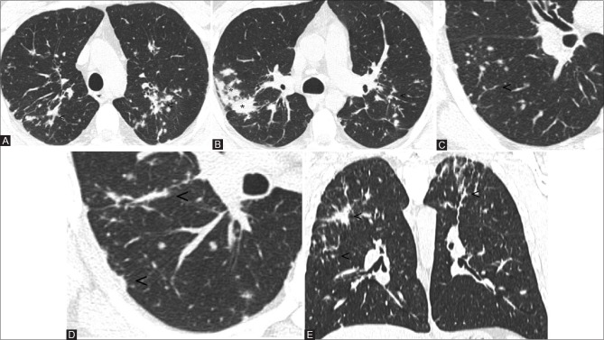
Axial images at diagnosis show bilateral lung nodules with a perilymphatic pattern. Note the peribronchovascular (A and B; see arrowheads), interlobular (C; see arrowheads), and subpleural involvement (D; see arrowheads). The confluence of many small nodules forms consolidations with irregular margins (B; see asterisk). The lesions mainly involve the upper and middle zones (E, coronal view; see arrowheads)
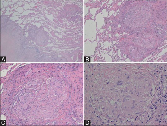
The thoracoscopic biopsy shows an interstitial fibrotic lesion with a reticular and nodular pattern (A, H and E stain, magnification 40×) and a subpleural and septal distribution. The lesion is characterized by multiple non-confluent granulomas (B, 100×) accompanied by collagen deposition devoid of significant inflammation and necrosis (C, 200×). The granulomas is made of epitheliod hystiocytes with few multinuclear giant cells (D, 400×)
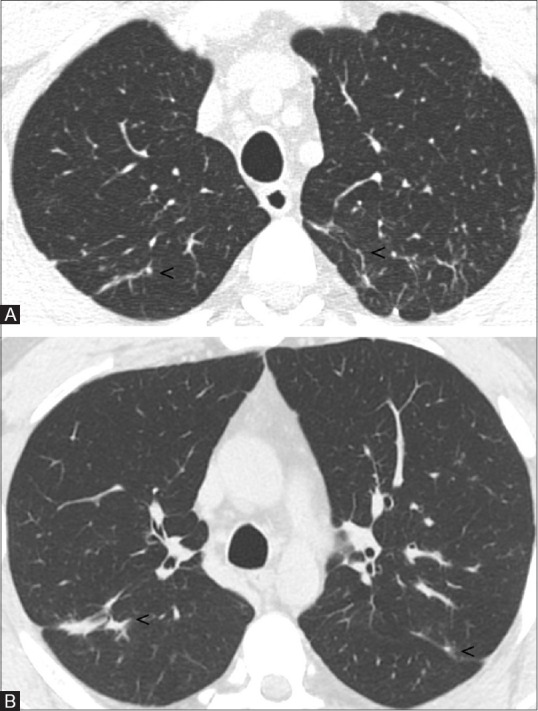
Two years later follow-up HRCT images. Bilaterally, some small linear opacities persist (A and B; see arrowheads), no further parenchymal abnormalities are appreciable
Similar articles
-
Hard Metal Lung Disease with Favorable Response to Corticosteroid Treatment: A Case Report and Literature Review.Tohoku J Exp Med. 2019 Jan;247(1):51-58. doi: 10.1620/tjem.247.51. Tohoku J Exp Med. 2019. PMID: 30674737 Review.
-
Hard metal lung disease: high resolution CT and histologic correlation of the initial findings and demonstration of interval improvement.J Thorac Imaging. 2005 Nov;20(4):301-4. doi: 10.1097/01.rti.0000181523.87391.a9. J Thorac Imaging. 2005. PMID: 16282911
-
Two-dimensional analysis of elements and mononuclear cells in hard metal lung disease.Am J Respir Crit Care Med. 2007 Jul 1;176(1):70-7. doi: 10.1164/rccm.200601-134OC. Epub 2007 Mar 15. Am J Respir Crit Care Med. 2007. PMID: 17363774
-
[Giant cell interstitial pneumonia associated with hard metals: a case report and review of the literature].Zhonghua Jie He He Hu Xi Za Zhi. 2009 Jul;32(7):493-6. Zhonghua Jie He He Hu Xi Za Zhi. 2009. PMID: 19954001 Review. Chinese.
-
Giant cell interstitial pneumonia in patients without hard metal exposure: analysis of 3 cases and review of the literature.Hum Pathol. 2016 Apr;50:176-82. doi: 10.1016/j.humpath.2015.12.004. Epub 2015 Dec 18. Hum Pathol. 2016. PMID: 26997453 Review.
Cited by
-
A comprehensive summary of disease variants implicated in metal allergy.J Toxicol Environ Health B Crit Rev. 2022 Aug 18;25(6):279-341. doi: 10.1080/10937404.2022.2104981. Epub 2022 Aug 16. J Toxicol Environ Health B Crit Rev. 2022. PMID: 35975293 Free PMC article. Review.
References
-
- Kim KI, Kim CW, Lee MK, Lee KS, Park CK, Choi SJ, et al. Imaging of Occupational Lung Disease. RadioGraphics. 2001;21:1371–91. - PubMed
-
- Nemery B, Abraham JL. Hard Metal Lung Disease Still Hard to Understand. Am J Respir Crit Care Med. 2007;176:2–3. - PubMed
-
- Choi JW, Lee KS, Chung MP, Han J, Chung MJ, Park JS. Giant Cell Interstial Pneumonia: High Resolution CT and Pathologic Findings in Four Adult Patient. AJR Am J Roentgenol. 2005;184:268–72. - PubMed
LinkOut - more resources
Full Text Sources
Other Literature Sources