

Argon attenuates the emergence of secondary injury after traumatic brain injury within a 2-hour incubation period compared to desflurane: an in vitro study
- PMID: 28744361
- PMCID: PMC5510299
- DOI: 10.4103/2045-9912.208512
Argon attenuates the emergence of secondary injury after traumatic brain injury within a 2-hour incubation period compared to desflurane: an in vitro study
Erratum in
-
Correction: Argon attenuates the emergence of secondary injury after traumatic brain injury within a 2-hour incubation period compared to desflurane: an in vitro study.Med Gas Res. 2017 Oct 17;7(3):155. doi: 10.4103/2045-9912.215756. eCollection 2017 Jul-Sep. Med Gas Res. 2017. PMID: 29152208 Free PMC article.
Abstract
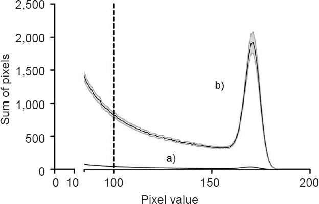
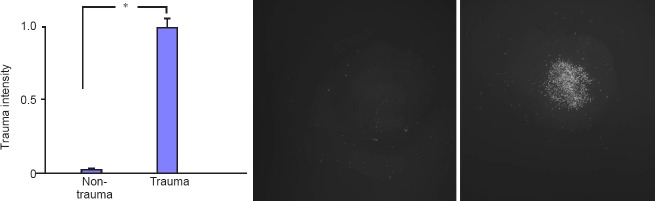
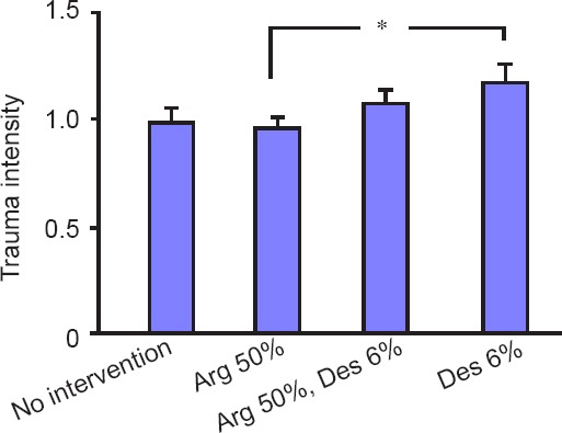
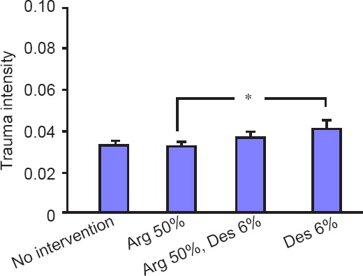
Despite years of research, treatment of traumatic brain injury (TBI) remains challenging. Considerable data exists that some volatile anesthetics might be neuroprotective. However, several studies have also revealed a rather neurotoxic profile of anesthetics. In this study, we investigated the effects of argon 50%, desflurane 6% and their combination in an in vitro TBI model with incubation times similar to narcotic time slots in a daily clinical routine. Organotypic hippocampal brain slices of 5- to 7-day-old mice were cultivated for 14 days before TBI was performed. Slices were eventually incubated for 2 hours in an atmosphere containing no anesthetic gas, argon 50% or desflurane 6% or both. Trauma intensity was evaluated via fluorescent imagery. Our results show that neither argon 50% nor desflurane 6% nor their combination could significantly reduce the trauma intensity in comparison to the standard atmosphere. However, in comparison to desflurane 6%, argon 50% displayed a rather neuroprotective profile within the first 2 hours after a focal mechanical trauma (P = 0.015). A 2-hour incubation in an atmosphere containing both gases, argon 50% and desflurane 6%, did not result in significant effects in comparison to the argon 50% group or the desflurane 6% group. Our findings demonstrate that within a 2-hour incubation time neither argon nor desflurane could affect propidium iodide-detectable cell death in an in vitro TBI model in comparison to the standard atmosphere, although cell death was less with argon 50% than with desflurane 6%. The results show that within this short time period processes concerning the development of secondary injury are already taking place and may be manipulated by argon.
Keywords: argon; desflurane; in vitro model; neuroprotection; organotypic hippocampal brain slices; propidium iodide; secondary injury; traumatic brain injury.
Conflict of interest statement
Conflicts of interest None declared.
Figures
References
-
- TBI care. Evidence-based Diagnostic and Treatment Planning for Traumatic Brain Injuries. [2016-08-12]. http://www.tbicare.eu/5 .
-
- World Health Organization. Neurological Disorders: Public Health Challenges. Geneva, Switzerland: World Health Organization; 2006.
-
- Finkelstein E, Corso PS, Miller TR. The Incidence and Economic Burden of Injuries in the United States. New York: Oxford University Press; 2006.
-
- Gustavsson A, Svensson M, Jacobi F, et al. Cost of disorders of the brain in Europe 2010. Eur Neuropsychopharmacol. 2011;21:718–779. - PubMed
-
- Olesen J, Gustavsson A, Svensson M, Wittchen HU, Jonsson B. The economic cost of brain disorders in Europe. Eur J Neurol. 2012;19:155–162. - PubMed
LinkOut - more resources
Full Text Sources
Other Literature Sources
