Review
. 2017 Jul 11:5:65.
doi: 10.3389/fcell.2017.00065. eCollection 2017.

New Challenges to Study Heterogeneity in Cancer Redox Metabolism

Affiliations
Review

New Challenges to Study Heterogeneity in Cancer Redox Metabolism

Rui Benfeitas et al. Front Cell Dev Biol. .

Abstract

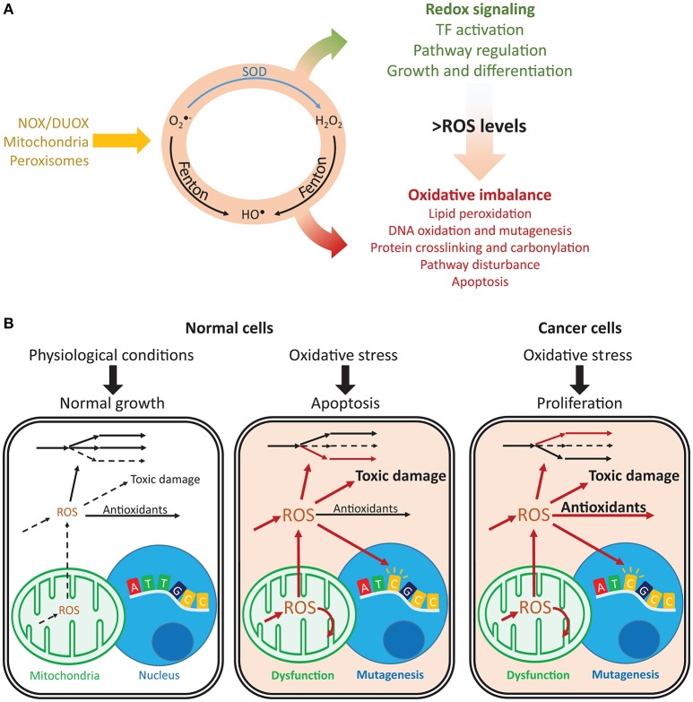
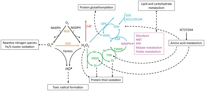
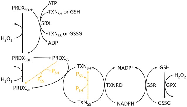
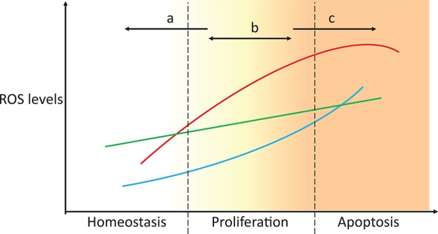
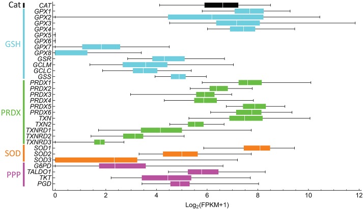
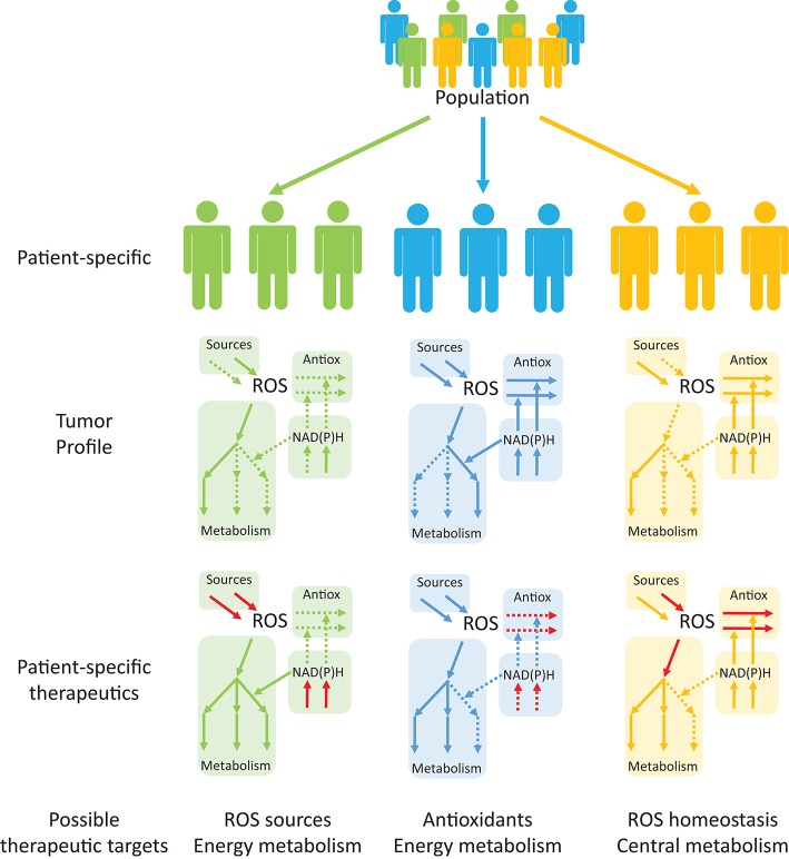
Reactive oxygen species (ROS) are important pathophysiological molecules involved in vital cellular processes. They are extremely harmful at high concentrations because they promote the generation of radicals and the oxidation of lipids, proteins, and nucleic acids, which can result in apoptosis. An imbalance of ROS and a disturbance of redox homeostasis are now recognized as a hallmark of complex diseases. Considering that ROS levels are significantly increased in cancer cells due to mitochondrial dysfunction, ROS metabolism has been targeted for the development of efficient treatment strategies, and antioxidants are used as potential chemotherapeutic drugs. However, initial ROS-focused clinical trials in which antioxidants were supplemented to patients provided inconsistent results, i.e., improved treatment or increased malignancy. These different outcomes may result from the highly heterogeneous redox responses of tumors in different patients. Hence, population-based treatment strategies are unsuitable and patient-tailored therapeutic approaches are required for the effective treatment of patients. Moreover, due to the crosstalk between ROS, reducing equivalents [e.g., NAD(P)H] and central metabolism, which is heterogeneous in cancer, finding the best therapeutic target requires the consideration of system-wide approaches that are capable of capturing the complex alterations observed in all of the associated pathways. Systems biology and engineering approaches may be employed to overcome these challenges, together with tools developed in personalized medicine. However, ROS- and redox-based therapies have yet to be addressed by these methodologies in the context of disease treatment. Here, we review the role of ROS and their coupled redox partners in tumorigenesis. Specifically, we highlight some of the challenges in understanding the role of hydrogen peroxide (H2O2), one of the most important ROS in pathophysiology in the progression of cancer. We also discuss its interplay with antioxidant defenses, such as the coupled peroxiredoxin/thioredoxin and glutathione/glutathione peroxidase systems, and its reducing equivalent metabolism. Finally, we highlight the need for system-level and patient-tailored approaches to clarify the roles of these systems and identify therapeutic targets through the use of the tools developed in personalized medicine.

Keywords: cancer heterogeneity; personalized medicine; reactive oxygen species; redox biology; systems biology.


Figures

Figure 1
Figure 1
Imbalances in ROS and redox cycles lead to contrasting outcomes in normal and cancer cells. (A) ROS are intracellularly produced by NADPH oxidases and dual oxidases (NOX/DUOX) through mitochondrial oxidative phosphorylation and in peroxisomes. Their interconversion (orange circle) occurs through enzyme catalyzed and non-catalyzed reactions. For instance, metal-catalyzed Fenton reactions produce HO from O2- and H2O2. Under low ROS levels, these oxidants control important signaling reactions, activating transcription factors, regulating pathways and controlling cell growth and differentiation. Under high ROS levels, oxidation of lipids, nucleic acids, and proteins is toxic and may disturb pathways and lead to cell death. (B) Normal and cancer cells present important differences in their responses to oxidative stress. Under normal conditions, ROS production is low and antioxidant defenses are sufficient to prevent toxic damage. Under oxidative stress, the promoted production of ROS overcomes the cell's capacity for detoxification and results in increased toxic damage and pathway disruption, which may lead to mitochondrial dysfunction, mutagenesis and ultimately apoptosis. In turn, cancer cells are under constant oxidative stress, which through upregulation of antioxidant defenses, prevents apoptosis while maintaining ROS toxicity. Arrows indicate fluxes, increasing from dashed to continuous, in red. O2-, superoxide; H2O2, hydrogen peroxide; HO, hydroxyl radical; SOD, superoxide dismutase.
Figure 2
Figure 2
Reactions involving ROS, antioxidant systems and energy metabolism. O2- and H2O2 are produced from oxygen through reactions that may oxidize NADPH (dotted arrows, e.g., catalysis by NADPH oxidases). O2- is dismutated to H2O2 by SOD, and O2- and H2O2 may be converted to HO by Fenton reactions. CAT, PRDX and GPX scavenge H2O2. The catalytic cycles of PRDX and TXN are represented, where SH and SS, respectively, indicate reduced and oxidized (disulfide) thiols. Boxes and dashed arrows indicate the external processes with which the metabolites are associated. For example, XCT/CD44 mediates cysteine import, which may then be incorporated into proteins, such as TXN and PRDX, or may be metabolized to yield GSH. Colors indicate proteins or processes from the same pathway.
Figure 3
Figure 3
Chemical cycle of PRDXs, TXNs, and GSH. The redox state of PRDXs and TXN is indicated as follows: SH, reduced cysteine thiol; SOH, sulfenic acid; SO2H, sulfinic acid; SS, disulfide. Orange dashed reactions highlight disulfide exchange between peroxiredoxins and thioredoxins and other proteins.
Figure 4
Figure 4
Targeting ROS homeostasis as a strategy for changing cell fate. In proliferative conditions, such as cancer, targeting antioxidant systems could move the redox state of the cell to either promote normal redox homeostasis or apoptosis (strategies a and c, respectively). Unsuccessful tackling of antioxidant metabolism results in the cells maintaining a proliferative state, which potentially enhances malignancy. The three lines represent the high heterogeneity between individuals, tissues, and cancer types.
Figure 5
Figure 5
Antioxidant gene expression greatly varies between liver hepatocellular carcinoma in different subjects. The gene expression of 50 subjects was downloaded from NCI's Genomic Data Commons, and fragments per kilobase transcript per million (FPKM) were computed. FPKM-values lower than one were considered to be non-expressed and were assigned a value of 0. The Log2(FPKM + 1) were then computed. Bars are colored according to processes of the same pathway, as indicated on the left. Genes and respective proteins: CAT, catalase; GPX1-8, glutathione peroxidase; GSR, glutathione reductase; GCLM and GCLC, glutamate-cysteine ligase modifier and catalytic subunits, respectively; GSS, glutathione synthetase; PRDX1-6, peroxiredoxin; TXN and TXN2, thioredoxin; TXNRD1-3, thioredoxin reductase; SOD, superoxide dismutase; G6PD, glucose-6-phosphate dehydrogenase; TALDO1, transaldolase 1, TKT, transketolase, and PGD, 6-phosphogluconate dehydrogenase.
Figure 6
Figure 6
Personalized systems medicine approaches are emerging as useful tools in devising patient-specific, rather than population-based, therapeutic targets in cancer. Tumor profiling of patients may help in identifying up- and down-regulated pathways (continuous and dashed arrows, respectively) that are suitable for therapeutic targeting. Drug targeting of specific processes (red arrows), either to promote or inhibit the processes, will permit alterations in the consequences of redox processes in cancer and other diseases.

References

    1. Agren R., Bordel S., Mardinoglu A., Pornputtapong N., Nookaew I., Nielsen J. (2012). Reconstruction of genome-scale active metabolic networks for 69 human cell types and 16 cancer types using INIT. PLoS Comput. Biol. 8:e1002518. 10.1371/journal.pcbi.1002518 - DOI - PMC - PubMed
    1. Agren R., Mardinoglu A., Asplund A., Kampf C., Uhlen M., Nielsen J. (2014). Identification of anticancer drugs for hepatocellular carcinoma through personalized genome-scale metabolic modeling. Mol. Syst. Biol. 10, 1–13. 10.1002/msb.145122 - DOI - PMC - PubMed
    1. Aran D., Sirota M., Butte A. J. (2015). Systematic pan-cancer analysis of tumour purity. Nat. Commun. 6:8971. 10.1038/ncomms9971 - DOI - PMC - PubMed
    1. Arnér E. S. J., Holmgren A. (2006). The thioredoxin system in cancer. Semin. Cancer Biol. 16, 420–426. 10.1016/j.semcancer.2006.10.009 - DOI - PubMed
    1. Arnold R. S., Shi J., Murad E., Whalen A. M., Sun C. Q., Polavarapu R., et al. . (2001). Hydrogen peroxide mediates the cell growth and transformation caused by the mitogenic oxidase Nox1. Proc. Natl. Acad. Sci. U.S.A. 98, 5550–5555. 10.1073/pnas.101505898 - DOI - PMC - PubMed