. 2018 Sep;68(5):555-565.
doi: 10.1007/s12576-017-0561-9. Epub 2017 Jul 25.

Xanthoceraside modulates neurogenesis to ameliorate cognitive impairment in APP/PS1 transgenic mice

Affiliations

Xanthoceraside modulates neurogenesis to ameliorate cognitive impairment in APP/PS1 transgenic mice

Lin Zhu et al. J Physiol Sci. 2018 Sep.

Abstract

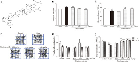
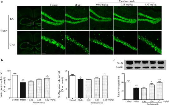
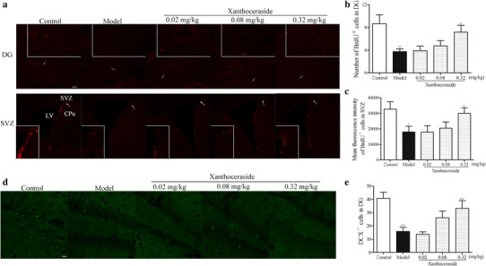
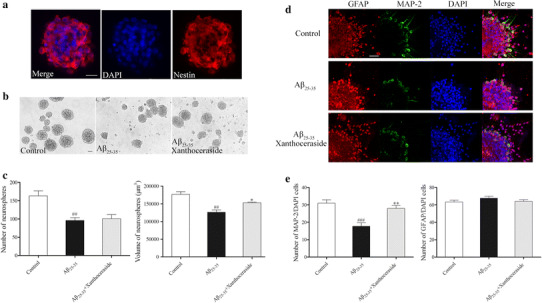
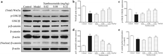
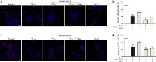
Neuronal loss is reported to be an important pathological process in Alzheimer's disease (AD). Neurogenesis is a process of generation of new neurons to fill the neuronal loss. Xanthoceraside has been shown to attenuate the cognitive deficits in several AD animal models. However, little is known about the effect of xanthoceraside on neurogenesis in APP/PS1 transgenic mice. Thus, in this study, we investigated whether xanthoceraside can ameliorate learning and memory impairment by promoting NSCs proliferation and neuronal differentiation. The results suggested that xanthoceraside significantly ameliorated the cognitive impairment and induced NSCs proliferation and neuronal differentiation in APP/PS1 transgenic mice. Meanwhile, in vitro study revealed that xanthoceraside increased the size of NSCs and induced NSCs differentiation into neurons compared with amyloid beta-peptide (25-35) (Aβ25-35) treatment. Furthermore, we found that xanthoceraside significantly increased the expression of Wnt3a and p-GSK3β, decreased the expression of p-β-catenin, and induced nuclear translocation of β-catenin in APP/PS1 transgenic mice. Furthermore, in vitro study found that the effect of xanthoceraside on inducing NSCs proliferation and neuronal differentiation were inhibited by Wnt pathway inhibitor Dickkopf-1 (Dkk-1). Our data demonstrated that xanthoceraside may promote the proliferation and differentiation of NSCs into neurons by up-regulating the Wnt/β-catenin pathway to fill the neuronal loss, thereby improving learning and memory impairment in APP/PS1 transgenic mice.

Keywords: Alzheimer’s disease; Learning and memory; Neurogenesis; Wnt/β-catenin pathway; Xanthoceraside.


Conflict of interest statement

The authors have no conflicts of interest to declare.

Figures

Fig. 1
Fig. 1
Effects of xanthoceraside on learning and memory impairment in APP/PS1 transgenic mice. a Chemical structure of xanthoceraside. b The pathway traveled by the mice during the open field test. c, d Total number of arm entries and spontaneous alternation behavior (%) in the Y-maze test. e, f Total exploration time and preferential index at 1 and 24 h during the testing phase. Values are expressed as the mean ± SEM (n = 11–12). ## p < 0.01 and ### p < 0.001 vs. control; **p < 0.01 and ***p < 0.001 vs. model
Fig. 2
Fig. 2
Effects of xanthoceraside on neurons in hippocampal DG and CA1 of APP/PS1 transgenic mice. a Immunostaining against the NeuN in hippocampal DG and CA1 regions. b Quantitative analysis of neurons in hippocampal DG and CA1 regions. Values are expressed as the mean ± SEM (n = 6). c Western blots showing the protein level of NeuN in the hippocampus. Scale bar = 20 μm. Values are expressed as the mean ± SEM (n = 5–6). # p < 0.05, ## p < 0.01 and ### p < 0.001 vs. control; *p < 0.05 and **p < 0.01 vs. model
Fig. 3
Fig. 3
Effects of xanthoceraside on proliferation and differentiation of NSCs in hippocampal DG and SVZ regions. a Confocal photomicrographs showing BrdU+ cells in hippocampal DG and SVZ regions. b, c Bar diagram showing the quantitative analysis of BrdU+ cells in hippocampus DG and SVZ regions. Scale bar = 100 μm. d, e Images and quantitative analysis of DCX+ cells in hippocampal DG region. Scale bar = 20 μm. Values are expressed as the mean ± SEM (n = 6). # p < 0.05, ## p < 0.01 and ### p < 0.001 vs. control; *p < 0.05 and **p < 0.01 vs. model
Fig. 4
Fig. 4
Effects of xanthoceraside on proliferation and differentiation of the hippocampus-derived NSCs in vitro. a Identification of neurospheres that were fixed and stained with NSCs marker nestin (red) and then counterstained with DAPI (blue) as the nuclear marker. b Neurosphere growth kinetics in the control, Aβ25–35 (20 μM), and Aβ25–35 (20 μM) combined with the xanthoceraside (0.1 μM)-treated group were studied by phase-contrast photomicrograph. c Bar diagram showing the number and volume of NSCs-derived neurospheres (neurospheres with a diameter ≥30 μm were used for calculating). d Images showing the effect of xanthoceraside on the differentiation potential of NSCs, as assessed by immuno-labeling with a neuronal marker (MAP-2, green) and an astrocyte marker (GFAP, red) and then counterstained with DAPI (blue). e A number of MAP-2-labeled and GFAP-labeled cells were calculated. Scale bar = 20 μm. Values are expressed as the mean ± SEM (n = 3). ## p < 0.01 and ### p < 0.001 vs. control; *p < 0.05 and **p < 0.01 vs. Aβ25–35 group
Fig. 5
Fig. 5
Effects of xanthoceraside on the Wnt/β-catenin signaling pathway in the hippocampus of APP/PS1 transgenic mice. a Representative image of immunoblots for Wnt3a, GSK3β, p-GSK3β, β-catenin, and p-β-catenin in the hippocampus. bd Quantitative analysis of Wnt3a, p-GSK3β, and p-β-catenin protein expression by Western blot. e Quantitative analysis of nuclear translocation of β-catenin by Western blot. Values are expressed as the mean ± SEM (n = 5–6). ## p < 0.01 and ### p < 0.001 vs. control; *p < 0.05, **p < 0.01 and ***p < 0.001 vs. model
Fig. 6
Fig. 6
Inhibition of the Wnt pathway affects xanthoceraside-mediated stimulatory effects on NSCs proliferation and neuronal differentiation in culture. a, c Representative immunofluorescent images showed that NSCs culture was treated with xanthoceraside in the presence and absence of the Wnt pathway inhibitor Dkk-1 in order to understand the effects on proliferation and differentiation. Scale bar = 20 μm. b, d Quantitative analysis of the proliferation and differentiation of NSCs. Scale bar = 50 μm. Values are expressed as the mean ± SEM (n = 3). ### p < 0.001 vs. control; *p < 0.05 and **p < 0.01 vs. Aβ25–35; $$ p < 0.01 and $$$ p < 0.001 vs. Aβ25–35 + xanthoceraside

References

    1. Xia M, Yang L, Sun G, Qi S, Li B. Mechanism of depression as a risk factor in the development of Alzheimer’s disease: the function of AQP4 and the glymphatic system. Psychopharmacology (Berl) 2016;234(3):365–379. doi: 10.1007/s00213-016-4473-9. - DOI - PubMed
    1. Dong H, Csernansky CA, Martin MV, Bertchume A, Vallera D, Csernansky JG. Acetylcholinesterase inhibitors ameliorate behavioral deficits in the Tg2576 mouse model of Alzheimer’s disease. Psychopharmacology. 2005;181(1):145–152. doi: 10.1007/s00213-005-2230-6. - DOI - PMC - PubMed
    1. Li XY, Men WW, Zhu H, Lei JF, Zuo FX, Wang ZJ, et al. Age- and brain region-specific changes of glucose metabolic disorder, learning, and memory dysfunction in early Alzheimer’s disease assessed in APP/PS1 transgenic mice using 18F-FDG-PET. Int J Mol Sci. 2016;17(10):1707. doi: 10.3390/ijms17101707. - DOI - PMC - PubMed
    1. Jankowsky JL, Fadale DJ, Anderson J, Xu GM, Gonzales V, Jenkins NA, et al. Mutant presenilins specifically elevate the levels of the 42 residue beta-amyloid peptide in vivo: evidence for augmentation of a 42-specific gamma secretase. Hum Mol Genet. 2004;13(2):159–170. doi: 10.1093/hmg/ddh019. - DOI - PubMed
    1. Parthsarathy V, McClean PL, Holscher C, Taylor M, Tinker C, Jones G, et al. A novel retro-inverso peptide inhibitor reduces amyloid deposition, oxidation and inflammation and stimulates neurogenesis in the APPswe/PS1DeltaE9 mouse model of Alzheimer’s disease. PLoS One. 2013;8(1):e54769. doi: 10.1371/journal.pone.0054769. - DOI - PMC - PubMed

LinkOut - more resources