. 2017 Nov 15;141(10):2050-2061.
doi: 10.1002/ijc.30906. Epub 2017 Aug 7.

BOK displays cell death-independent tumor suppressor activity in non-small-cell lung carcinoma

Affiliations

BOK displays cell death-independent tumor suppressor activity in non-small-cell lung carcinoma

Erika Moravcikova et al. Int J Cancer. .

Abstract

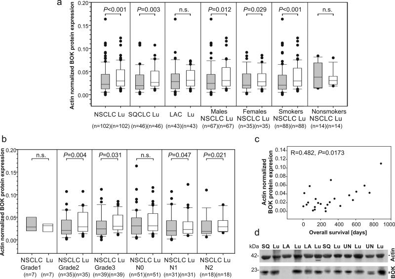
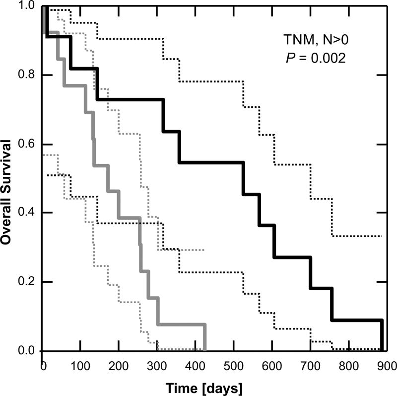
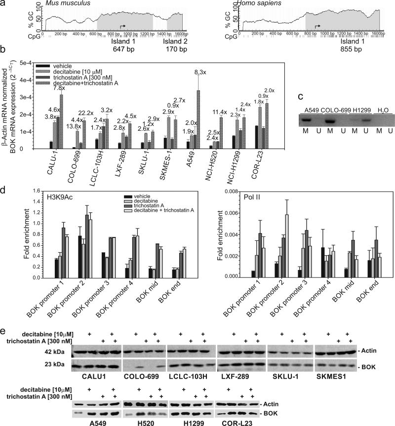
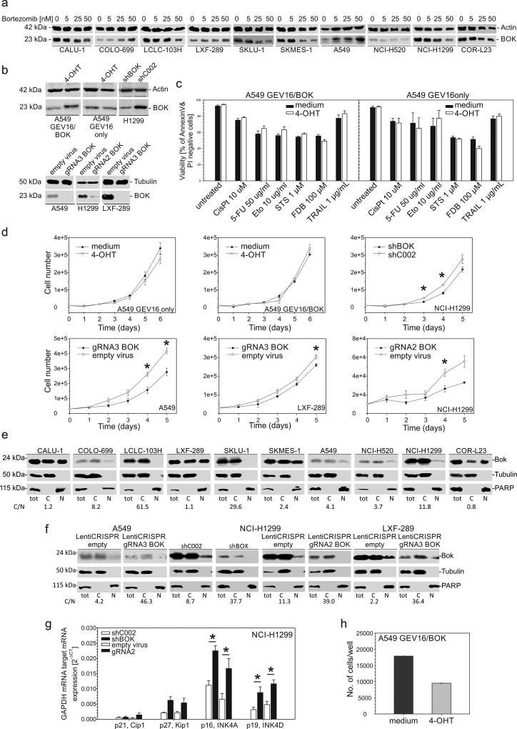
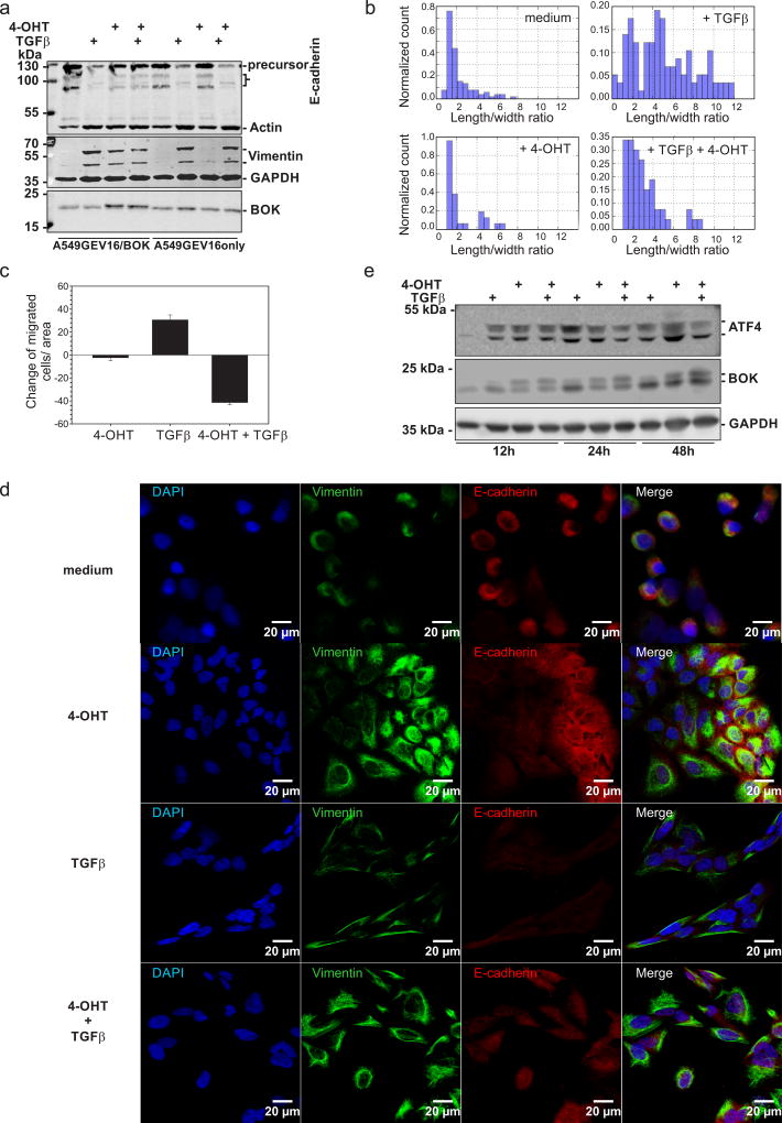
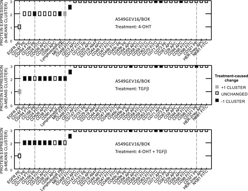
As the genomic region containing the Bcl-2-related ovarian killer (BOK) locus is frequently deleted in certain human cancers, BOK is hypothesized to have a tumor suppressor function. In the present study, we analyzed primary non-small-cell lung carcinoma (NSCLC) tumors and matched lung tissues from 102 surgically treated patients. We show that BOK protein levels are significantly downregulated in NSCLC tumors as compared to lung tissues (p < 0.001). In particular, we found BOK downregulation in NSCLC tumors of grades two (p = 0.004, n = 35) and three (p = 0.031, n = 39) as well as in tumors with metastases to hilar (pN1) (p = 0.047, n = 31) and mediastinal/subcarinal lymph nodes (pN2) (p = 0.021, n = 18) as opposed to grade one tumors (p = 0.688, n = 7) and tumors without lymph node metastases (p = 0.112, n = 51). Importantly, in lymph node-positive patients, BOK expression greater than the median value was associated with longer survival (p = 0.002, Mantel test). Using in vitro approaches, we provide evidence that BOK overexpression is inefficient in inducing apoptosis but that it inhibits TGFβ-induced migration and epithelial-to-mesenchymal transition (EMT) in lung adenocarcinoma-derived A549 cells. We have identified epigenetic mechanisms, in particular BOK promoter methylation, as an important means to silence BOK expression in NSCLC cells. Taken together, our data point toward a novel mechanism by which BOK acts as a tumor suppressor in NSCLC by inhibiting EMT. Consequently, the restoration of BOK levels in low-BOK-expressing tumors might favor the overall survival of NSCLC patients.

Keywords: BOK; Bcl-2 family; apoptosis; epithelial-to-mesenchymal transition; non-small-cell lung carcinoma.


Conflict of interest statement

Conflict of Interest

The authors declare no conflicts of interest

Figures

Figure 1
Figure 1
BOK protein expression in NSCLC tissues and adjacent lung tissue. (a and b) Expression data are shown as median with the upper ranges of 75% (box) and 90% (whisker) and the lower ranges of 25% (box) and 10% (whisker). Statistical differences were calculated by the Wilcoxon signed rank test. P values lower than 0.05 were considered statistically significant. (c) Spearman’s correlation between BOK protein expression in lymph node positive NSCLC and patients’ overall survival. (d) A representative image of quantitative western blot of total protein lysates from NSCLC and lung parenchyma. SQ - squamous cell lung carcinoma, LA - lung adenocarcinoma, UN- undifferentiated cell lung carcinoma, Lu - lung parenchyma.
Figure 2
Figure 2
BOK expression level is predictive of survival in lymph node positive patients. Kaplan-Meier survival analysis: dashed lines = 95% confidence intervals. P-value was calculated according to the Mantel test. Grey < median BOK, Black ≥ median BOK.
Figure 3
Figure 3
The BOK gene is epigenetically silenced in NSCLC cell lines. (a) Bioinformatic analysis of human BOK gene methylation. CpG island prediction was based on DNA regions > 100bp with a CG content > 0.5 and observed/expected CpG ratio above 0.6. An input sequence of 1600-base long DNA around the BOK transcriptional start (arrow) was searched. (b) Cells were treated as indicated with 10 µM of decitabine for 72h or 300 nM of trichostatin A for 24 h or a combination of both; mRNA expression was measured by RT-qPCR. Data represent mean ± S.E.M from 3 experiments. Fold change between control and treatment is indicated above the bar. (c) Methylation status of BOK promoter by methylation specific PCR in A549, COLO-699 and H1299 cells using primers for methylated (M) or unmethylated (U) bisulfite-modified DNA. Representative example from 3 independent experiments is shown. (d) Acetylated H3K9 active chromatin mark and RNA polymerase II binding throughout the BOK gene in A549 cells. A549 cells treated with decitabine (72h) and/or trichostatin A (24h) were analyzed by ChIP with anti-H3K9Ac, anti-Pol II and IgG, followed by qPCR analysis of amplicons in the promoter and in the body of the BOK gene (internal control). Error bars indicate the standard error of the mean (SEM) of two biological replicates. (e) NSCLC cells were treated as indicated in (b) and total protein lysates analyzed by western blotting.
Figure 4
Figure 4
Stabilization of BOK protein by bortezomib, cell death and proliferation analyses. (a) Cells were treated with bortezomib at the indicated concentration for 24 h; total protein lysates were analyzed by western blotting. (b) Western blot analysis of total protein lysates from A549GEV16/BOK and A549GEV16only cells treated with or without 100 nM 4-OHT for 48h and of H1299, A549 and LXF-289 cells with downregulated BOK using shRNA or CRISPR/Cas9 technology. (c) A549GEV16/BOK or A549GEV16only cells were pretreated with or without 100nM 4-OHT overnight and treated with cisplatin, 5-FU, etoposide, staurosporine, fludarabine or TRAIL for 48 h. Viability was assessed by FITC-AnnexinV/PI staining using flow cytometry. Data represent mean ± S.E.M. from 3–6 independent experiments. (d) Trypan-blue exclusion assay of cell proliferation. Data represent mean ± S.E.M. from 3 independent experiments. Statistical analysis was performed using Student’s t-test. Asterisks indicate a P value lower than 0.05. (e and f) Detection of nuclear BOK by western blot analysis. Nuclear and cytoplasmic fractions were prepared from NSCLC cells. The purity of fractions was confirmed by detection of PARP (nuclear) or Tubulin (cytoplasmic). The cytoplasmic/nuclear ratio of BOK protein expression (C/N) is shown below the blots. (g) mRNA expression of GAPDH-normalized cell cycle regulators by RT-qPCR. Data represent mean ± S.E.M. from 3 independent experiments. Asterisks indicate a P value lower than 0.05 analyzed by Student’s t-test. (h) Anchorage-independent growth of A549GEV16/BOK cells in soft agar for 7 days. Cells were treated as indicated with or without 100 nM 4-OHT.
Figure 5
Figure 5
BOK inhibits EMT. Cells were pretreated with or without 100 nM 4-OHT and treated with or without 10 ng/ml of TGFβ2 for 48 hours. (a) Whole cell lysates were analyzed by western blotting using anti-E-cadherin, anti-actin (loading control), anti-BOK (same membrane), anti-vimentin and anti-GAPDH (loading control) antibodies. (b) Histogram of morphological changes of A549GEV16/BOK cells: the length and width of at least 100 cells were measured per condition. (c) Boyden chamber migration assay (d) Confocal scanning micrographs of A549GEV16/BOK cells. Anti-vimentin (green) and anti-E-cadherin (red) antibodies were used. All cells were counterstained with DAPI (blue). The same exposure times and LUT settings were used. (e) Western blot analysis of A549GEV16/BOK whole cell lysates treated as indicated above for 12, 24 and 48 hours. Membrane was probed with anti-ATF4, anti-BOK and anti-GAPDH (loading control) antibodies. One representative immunoblot is shown from three independent experiments.
Figure 6
Figure 6
Cell surface protein expression by flow cytometry. Thirty-eight markers shown were selected from 238 measured specificities on the basis of strength of expression or extent of modulation. Cells were treated as indicated with or without 100 nM 4-OHT overnight, following addition of 10 ng/ml of TGFβ or vehicle and incubated for 48 h. K-means cluster 0: no expression, 1: low expression, 2: moderate expression, 3: high expression. Y axis: Cell surface marker expression in untreated A549GEV16/BOK. Color coded boxes indicate changes between treatments (4-OHT and/or TGFβ) and control cells (vehicle-treated). Dashed lines indicate markers that were modulated by BOK overexpression.

References

    1. Llambi F, Wang YM, Victor B, Yang M, Schneider DM, Gingras S, Parsons MJ, Zheng JH, Brown SA, Pelletier S, Moldoveanu T, Chen T, et al. BOK Is a Non-canonical BCL-2 Family Effector of Apoptosis Regulated by ER-Associated Degradation. Cell. 2016;165(2):421–33. - PMC - PubMed
    1. Echeverry N, Bachmann D, Ke F, Strasser A, Simon HU, Kaufmann T. Intracellular localization of the BCL-2 family member BOK and functional implications. Cell Death Differ. 2013;20(6):785–99. - PMC - PubMed
    1. Hsu SY, Kaipia A, McGee E, Lomeli M, Hsueh AJ. Bok is a pro-apoptotic Bcl-2 protein with restricted expression in reproductive tissues and heterodimerizes with selective anti-apoptotic Bcl-2 family members. Proc Natl Acad Sci U S A. 1997;94(23):12401–6. - PMC - PubMed
    1. Inohara N, Ekhterae D, Garcia I, Carrio R, Merino J, Merry A, Chen S, Nunez G. Mtd, a novel Bcl-2 family member activates apoptosis in the absence of heterodimerization with Bcl-2 and Bcl-XL. J Biol Chem. 1998;273(15):8705–10. - PubMed
    1. Einsele-Scholz S, Malmsheimer S, Bertram K, Stehle D, Johanning J, Manz M, Daniel PT, Gillissen BF, Schulze-Osthoff K, Essmann F. Bok is a genuine multi-BH-domain protein that triggers apoptosis in the absence of Bax and Bak. J Cell Sci. 2016;129(11):2213–23. - PubMed

Publication types

MeSH terms