. 2017 Aug 24;121(33):7899-7906.
doi: 10.1021/acs.jpcb.7b05568. Epub 2017 Aug 9.

Nanoscale Ion Pump Derived from a Biological Water Channel

Affiliations

Nanoscale Ion Pump Derived from a Biological Water Channel

Karl Decker et al. J Phys Chem B. .

Abstract

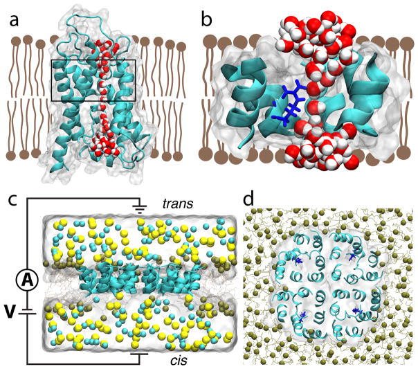
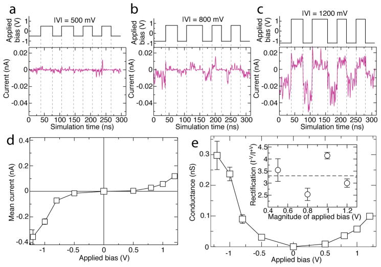
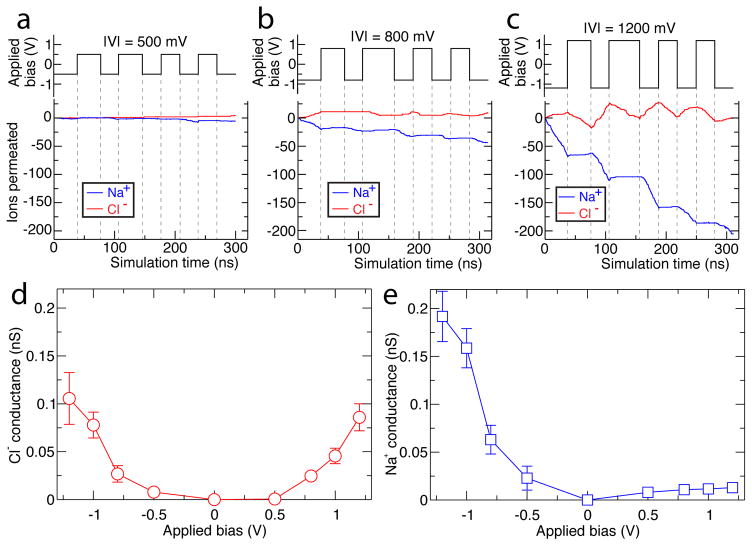
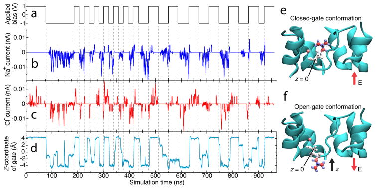
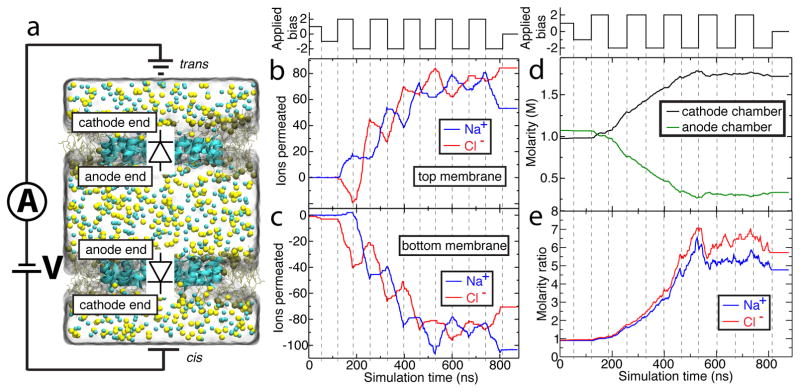
Biological molecular machines perform the work of supporting life at the smallest of scales, including the work of shuttling ions across cell boundaries and against chemical gradients. Systems of artificial channels at the nanoscale can likewise control ionic concentration by way of ionic current rectification, species selectivity, and voltage gating mechanisms. Here, we theoretically show that a voltage-gated, ion species-selective, and rectifying ion channel can be built using the components of a biological water channel aquaporin. Through all-atom molecular dynamics simulations, we show that the ionic conductance of a truncated aquaporin channel nonlinearly increases with the bias magnitude, depends on the channel's orientation, and is highly cation specific but only for one polarity of the transmembrane bias. Further, we show that such an unusually complex response of the channel to transmembrane bias arises from mechanical motion of a positively charged gate that blocks cation transport. By combining two truncated aquaporins, we demonstrate a molecular system that pumps ions against their chemical gradients when subject to an alternating transmembrane bias. Our work sets the stage for future biomimicry efforts directed toward reproducing the function of biological ion pumps using synthetic components.


Figures

Figure 1
Figure 1
All-atom model of truncated aquaporin. (a,b) Side-on view of wild-type aquaporin monomer (panel a) and its truncated version (panel b). The secondary structure of the AQP proteins is shown using a cartoon representation; each protein’s boundaries are depicted by a semi-transparent molecular surface. The lipid bilayer is shown as brown schematic, and the water wire through the AQP monomers is shown using red (oxygen) and white (hydrogen) spheres. In panel a, the region to be left after truncation of the protein is indicated by a black rectangle. In panel b, residue Arg-197 is additionally shown in blue licorice. To provide a better view of the water wire, certain residues of the proteins are not shown. (c,d) Side (panel c) and top (panel d) views of the system used to characterize ion conductance of truncated AQP. Ochre lines and spheres illustrate the truncated POPE membrane (spheres represent the phosphorous atoms of lipids); the semi-transparent surface illustrates the volume occupied by 1 M NaCl electrolyte; Na+ and Cl ions are depicted as yellow and cyan spheres, respectively. For clarity, electrolyte is not shown in the top view of the system. The circuit diagram in panel c indicates applied bias and measurement of transmembrane ionic current.
Figure 2
Figure 2
Ionic current, conductance, and rectification of truncated AQP system. (a–c) Ionic current across truncated AQP embedded in a lipid membrane, Figure 1c,d, at ±500 mV, ±800 mV, and ±1200 mV biases respectively, with magnitude of bias given as —V— at the top of each panel. Data at ±1000 mV are reported in Figure S1. Purple traces indicate current whereas step-function black traces indicate the applied transmembrane bias versus simulation time. The ionic current traces show 2 ns running average of the instantaneous current sampled every 9.6 ps. Dashed lines serve as guides for the eye. Positive current travels from cis to trans as defined in Figure 1c. (d,e) Mean ionic current (d) and ion conductance (e) of the truncated AQP tetramer as a function of transmembrane bias. Mean current is calculated by, first, finding the time-average of the ionic current across the tetramer for each constant bias fragment of the trajectory. The weighted average of these currents is the mean current reported. Error bars in both panels indicate the standard deviation of the mean. The mean conductance and its standard deviation are found by dividing mean current by magnitude of applied bias. The inset of (e) shows the ratios of the current at negative biases to the current at the corresponding positive biases. Error bars specify the propagated standard deviations in the mean currents.
Figure 3
Figure 3
Species-specific ionic conductance of truncated AQP. (a–c) The number of ions permeated across the truncated AQP tetramer system at ±500 mV, ±800 mV, and ±1200 mV biases respectively, with magnitude of bias given as —V— at the top of each panel. Red traces indicate Cl permeation, blue traces indicate Na+ permeation. Step-function black traces indicate the applied transmembrane bias versus simulation time, with dashed lines serving as guides for the eye. Data at ±1000 mV are reported in Figure S1. (d–e) Mean Cl (d) and Na+ (e) conductance of the truncated AQP tetramer. Error bars indicate the standard deviation of the mean conductance. The mean values were computed as described in the caption to Figure 2.
Figure 4
Figure 4
Mechanism of species-specific gating in truncated AQP. (a–d) Correlation of applied bias (a) and Na+ (b) and Cl (c) currents through one of the monomers of the truncated AQP tetramer, compared with displacement of that monomer’s Arg-197 side chain (d). In panel d, the location of the Arg-197 side chain is characterized by the z-coordinate of the chain’s guanadine group. Dashed lines serve as guides to the eye. The z axis is defined in panels e and f. (e,f) Molecular mechanism of truncated AQP gating. The terminal carbon in the positively charged guanadine group of Arg-197 is pictured in blue with a “+” on it. Arg-197, the residue responsible for gating, is rendered as spheres and sticks colored according to individual atomic charge (blue for positive, red for negative). At positive bias (panel e), the channel is closed, at negative bias (panel f) it is open. The direction of electric field is shown with red arrows, the direction of the z-axis is shown with a black arrow, and the location of the carbon atom whose position constitutes z = 0 is labeled. The truncated AQP is shown in cyan; water, ions, lipids, and one protein alpha-helix are omitted for clarity.
Figure 5
Figure 5
Performance of an ion pump built using truncated AQP. (a) Simulation system with parts depicted as in Figure 1c. In this double-membrane system, the bottom truncated AQP tetramer is oriented as in Figure 1c, while the top complex faces opposite. Because of the opposing orientations, the gate residues of one tetramer remain closed while those of the other are open. Note that the use of periodic boundary conditions means that the upper and lower partitions depicted here function as single chamber, referred to as the cathode chamber. (b–c) The number of ions permeated through the top (b) and bottom (c) truncated AQP tetramers of the double-membrane system as a function of the simulation time. The step-function black trace above panel b indicates the applied bias. Ion permeation across either membrane in the cis to trans direction (defined in panel a) increases the ion count; transport in the opposite direction decreases it. Na+ overwhelmingly travels from the anode (center) chamber to the cathode (outer two partitions) chamber in response to applied bias for as long as the gates remain effective. Dotted lines serve as guides for the eye. (d) Molarity of NaCl in the anode (green) and cathode (black) chambers. The step-function black trace above panel d indicates the applied bias. (e) Ratio of Na+ (blue trace) and Cl (red trace) molarity in the cathode chamber to that of the anode chamber.

Similar articles

Cited by

References

    1. van den Heuvel MGL, Dekker C. Motor Proteins at Work for Nanotechnology. Science. 2007;317:333–336. - PubMed
    1. Will CL, Lührmann R. Spliceosome Structure and Function. Cold Spring Harb Perspect Biol. 2011:3. - PMC - PubMed
    1. Schrempf H, OS, Kümmerlen R, Hinnah S, Müller D, Betzler M, Steinkamp T, Wagner R. A Prokaryotic Potassium Ion Channel with Two Predicted Transmembrane Segments from Streptomyces Lividans. EMBO J. 1995;14:5170–5178. - PMC - PubMed
    1. Bachand GD, Montemagno CD. Constructing Organic/Inorganic NEMS Devices Powered by Biomolecular Motors. Biomedical Microdevices. 2000;2:179–184.
    1. Soong RK, Bachand GD, Neves HP, Olkhovets AG, Craighead HG, Montemagno CD. Powering an Inorganic Nanodevice with a Biomolecular Motor. Science. 2000;290:1555–1558. - PubMed

Publication types

LinkOut - more resources