. 2017 Dec;56(6):1119-1127.
doi: 10.1002/mus.25752. Epub 2017 Aug 13.

Osteopontin is linked with AKT, FoxO1, and myostatin in skeletal muscle cells

Affiliations

Osteopontin is linked with AKT, FoxO1, and myostatin in skeletal muscle cells

Peter P Nghiem et al. Muscle Nerve. 2017 Dec.

Abstract

Introduction: Osteopontin (OPN) polymorphisms are associated with muscle size and modify disease progression in Duchenne muscular dystrophy (DMD). We hypothesized that OPN may share a molecular network with myostatin (MSTN).

Methods: Studies were conducted in the golden retriever (GRMD) and mdx mouse models of DMD. Follow-up in-vitro studies were employed in myogenic cells and the mdx mouse treated with recombinant mouse (rm) or human (Hu) OPN protein.

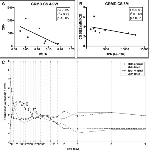
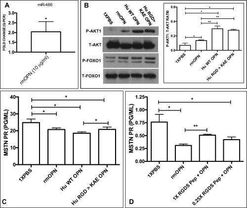
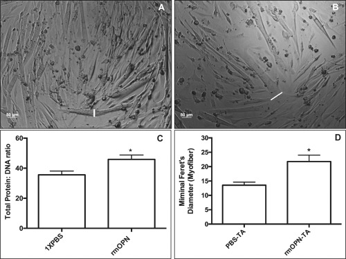
Results: OPN was increased and MSTN was decreased and levels correlated inversely in GRMD hypertrophied muscle. RM-OPN treatment led to induced AKT1 and FoxO1 phosphorylation, microRNA-486 modulation, and decreased MSTN. An AKT1 inhibitor blocked these effects, whereas an RGD-mutant OPN protein and an RGDS blocking peptide showed similar effects to the AKT inhibitor. RMOPN induced myotube hypertrophy and minimal Feret diameter in mdx muscle.

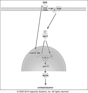
Discussion: OPN may interact with AKT1/MSTN/FoxO1 to modify normal and dystrophic muscle. Muscle Nerve 56: 1119-1127, 2017.

Keywords: AKT; Duchenne; GRMD; dog; mdx; muscle; myostatin; osteopontin.


Figures

Figure 1
Figure 1
OPN and MSTN were inversely correlated in GRMD dogs. (A) OPN and MSTN mRNA expression were inversely correlated in the CS at 4–9 weeks in GRMD dogs (r = –0.85, r 2 = 0.72; P < 0.05; n = 8), where myofiber hypertrophy is observed before gross hypertrophy. (B) OPN was inversely correlated with CS muscle circumference in GRMD dogs at 6 months of age (r = –0.83, r 2 = 0.69; P < 0.05; n = 8). (C) Cardiotoxin‐induced muscle injury in WT mice resulted in an immediate and substantial increase in OPN with a temporally concurrent reduction in MSTN expression at day 1 postinjection. OPN levels eventually returned to day 0 (pre‐injection) levels while MSTN returned to subnormal levels.
Figure 2
Figure 2
Recombinant mouse (rm)OPN treatment decreased MSTN expression. The rmOPN was dosed at 1, 5, and 10 μg/ml in H‐2kb‐tsA58 WT, conditionally immortalized myoblasts, and 10 μg/ml in myotubes. All mRNA and protein experiments were performed with 4–6 replicates. MSTN protein (PR) levels (pg/ml) were measured by enzyme‐linked immunoassay and normalized to total protein levels. Control (1× PBS) samples are shown in the far left column for panels (B)–(D) (*P ≤ 0.05; ***P ≤ 0.001). (A) rmOPN decreased endogenous MSTN mRNA in myoblasts in a dose‐dependent fashion at 24 hours. (B) rmOPN decreased MSTN protein in a dose‐dependent fashion in myoblasts at 48 hours. (C) rmOPN decreased MSTN protein in a dose‐dependent fashion in myoblasts at 48 hours. (D) rmOPN decreased MSTN protein in myotubes at 24 hours.
Figure 3
Figure 3
Recombinant mouse (rm)OPN treatment of H‐2kb‐tsA58 WT myoblasts led to phosphorylation of AKT1 and FoxO1. AKT1 and FoxO1 levels were measured by Western blot and quantified by densitometry. Phosphorylated AKT1 and FoxO1 were normalized to total AKT and FoxO1 levels, respectively. The numbers to the left of the blots indicate molecular weight in kilodaltons (*P < 0.05; **P < 0.01). T = total; P = phosphorylated. (A) AKT1 phosphorylation was induced in rmOPN‐treated (10 μg/ml) myogenic cells after 24 hours of incubation compared with control (1× PBS). The AKT inhibitor #124005 blocked this effect. FoxO1 phosphorylation was also greater in rmOPN‐treated cells compared with control, and AKT inhibitor also blocked this effect. Duplicate to quadruplicates were completed, as shown in each blot. Please note that all samples on each lane were run on the same gel. (B, C) Phosphorylated levels of AKT1 and FoxO1 were increased in rmOPN‐treated cells, whereas treatment with the AKT inhibitor decreased these levels to below control values.
Figure 4
Figure 4
MSTN and FoxO1 decreased with rmOPN treatment and this effect was rescued by an AKT inhibitor. (A) MSTN and FoxO1 mRNA were decreased in H‐2kb‐tsA58 WT, conditionally immortalized myoblasts after rmOPN treatment (10 μg/ml). Addition of AKT inhibitor #124005 rescued MSTN [fold‐change (FC) = +1.17] and FoxO1 (FC = +1.452) mRNA expression. (B) MSTN protein decreased after rmOPN treatment and was rescued with AKT inhibitor (FC = +1.4) in myoblasts.
Figure 5
Figure 5
AKT1 is activated by OPN through RGD‐ and non–RGD‐dependent receptors. Hu‐WT OPN (normal RGD sequence) and Hu‐RGD→KAE OPN (lacking the ITG‐binding RGD sequence) were tested in H‐2kb‐tsA58 WT myoblasts. (A) miR‐486 was increased in rmOPN‐treated myoblasts (FC = +2.07; P < 0.05) (*P ≤ 0.05; **P ≤ 0.01). (B) rmOPN, Hu‐RGD→KAE OPN, and Hu‐WT OPN increased AKT phosphorylation compared with 1× PBS control, with the latter showing the greatest effect (P < 0.05). Both human OPN proteins showed greater AKT1 phosphorylation abilities compared with rmOPN (P < 0.01). All OPN proteins increased FoxO1 phosphorylation. (C) Hu‐WT OPN, Hu‐RGD→KAE OPN, and rmOPN all decreased MSTN compared with 1× PBS control (P < 0.05). Hu‐WT OPN decreased MSTN protein slightly further compared with Hu‐RGD→KAE OPN (P < 0.05). (D) rmOPN decreased MSTN protein compared with 1× PBS (P < 0.05). This effect was partially blocked when rmOPN was co‐treated with an RGDS amino acid blocking peptide [0.05 (0.25×) and 0.2 mg/ml (1×)] compared with rmOPN alone, but was not dose‐dependent (P < 0.01).
Figure 6
Figure 6
Myotubes and muscle treated with rmOPN displayed hypertrophy. Myotubes were treated with rmOPN for 24–48 hours and subsequently evaluated on day 5 of differentiation. A 10‐μg/ml dose for rmOPN optimal and was used for each experiment. (A) Myotubes were treated with 1× PBS to serve as a control. (B) Several of the H‐2kb‐tsA58 WT myotubes exhibited increased cell diameter (white lines) compared with 1× PBS. (C) Treatment of myotubes with rmOPN increased total protein content by 26.6% after normalizing to total DNA content compared with control (P < 0.05). All experiments were performed in sextuplicates. (D) There was increased minimal Feret diameter in rmOPN‐injected tibialis anterior (TA) muscles of 4‐week‐old mdx mice compared with contralateral saline‐injected TA muscles. Mice were injected at 3 weeks of age (1‐week treatment duration). The minimal Feret diameter was measured only for myofibers with green dye immediately adjacent to the myofiber membrane. N = 4 limbs per group.
Figure 7
Figure 7
Schematic showing proposed pathway whereby OPN may interact with hypertrophy‐related proteins in muscle cells to induce hypertrophy. FoxO1 is a transcriptional regulator of MSTN, binding its promoter region to activate transcription. FoxO1 phosphorylation by AKT1 prevents translocation to the nucleus, thereby interfering with its transcriptional functions, including activation of MSTN. OPN also increased miR‐486, which could modulate FoxO1 mRNA and protein levels. We hypothesized that OPN binds integrins (ITG) and/or CD44 to activate AKT1, downregulate FoxO1, increase FoxO1 protein phosphorylation, and decrease MSTN mRNA and protein expression, resulting in myotube and myofiber hypertrophy.

References

    1. Scatena M, Liaw L, Giachelli CM. Osteopontin: a multifunctional molecule regulating chronic inflammation and vascular disease. Arterioscler Thromb Vasc Biol 2007;27:2302–2309. - PubMed
    1. Liaw L, Almeida M, Hart CE, Schwartz SM, Giachelli CM. Osteopontin promotes vascular cell adhesion and spreading and is chemotactic for smooth muscle cells in vitro. Circ Res 1994;74:214–224. - PubMed
    1. Urtasun R, Lopategi A, George J, Leung TM, Lu Y, Wang X, et al Osteopontin, an oxidant stress sensitive cytokine, up‐regulates collagen‐I via integrin α(V)β(3) engagement and PI3K/pAkt/NFκB signaling. Hepatology 2012;55:594–608. - PMC - PubMed
    1. Zheng DQ, Woodard AS, Tallini G, Languino LR. Substrate specificity of alpha(v)beta(3) integrin‐mediated cell migration and phosphatidylinositol 3‐kinase/AKT pathway activation. J Biol Chem 2000;275:24565–24574. - PubMed
    1. Von Marschall Z, Fisher LW. Dentin matrix protein‐1 isoforms promote differential cell attachment and migration. J Biol Chem 2008;283:32730–32740. - PMC - PubMed