

Review
doi: 10.1021/acs.biochem.7b00435.
Epub 2017 Jul 26.
Bioluminescent Probes for Imaging Biology beyond the Culture Dish
Affiliations
- PMID: 28745860
- PMCID: PMC5914164
- DOI: 10.1021/acs.biochem.7b00435
Item in Clipboard
Review
Bioluminescent Probes for Imaging Biology beyond the Culture Dish
Biochemistry.
.
Abstract
Bioluminescence with luciferase-luciferin pairs is an attractive method for surveying cells in live tissues and whole organisms. Recent advances in luciferin chemistry and luciferase engineering are further expanding the scope of the technology. It is now possible to spy on cells in a variety of deep tissues and visualize multicellular interactions, feats that are enabling new questions to be asked and new ideas to be explored. This perspective piece highlights recent successes in bioluminescent probe development and their applications to imaging in live cells, tissues, and animals.
Figures

A) D-Luciferin is oxidized by firefly luciferase (Fluc) to produce oxyluciferin and a photon of light. B) Coelenterazine is oxidized by a variety of marine luciferases, including Renilla luciferase (Rluc), Gaussia luciferase (Gluc), and Oplophorus luciferase (Oluc). These luciferases, unlike Fluc, require only oxygen as a cofactor in the light-emitting reaction.

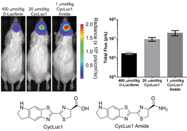
A) D-Luciferin and related analogs can be accessed using Appel’s salt and C-H activation chemistry. B) A diverse array of coelenterazine analogs has been synthesized in recent years.

Native luciferins are highlighted with colored boxes. Brackets denote the wavelength (nm) of maximum bioluminescence emission observed upon incubation of the compound with luciferase. While many analogs can provide unique colors of light, most are not efficiently processed by native luciferases.

A) Antares comprises a fusion of NanoLuc with two cyan-excitable orange fluorescent proteins (CyOFP1). B) The BRET fusion exhibits red-shifted emission relative to NanoLuc. ONL: Orange Nano-lantern. C) Antares enabled sensitive imaging with low luciferin levels in mice. Antares and Fluc genes were delivered to the liver via hydrodynamic injection. Luciferins were supplied intravenously. Adapted with permission from ref .
(Left) Luciferase expression in brain was achieved via viral transduction. Luciferins were via administered intraperitoneal injection. (Right) Photon flux from mouse brains pictured. Adapted with permission from ref .

A) A strategy for multicomponent bioluminescence imaging with luciferin analogs and mutant luciferases. B) The 4′ and 7′ sites of D-luciferin were targeted to preclude binding to native Fluc. C) Substrate resolution was achieved in mouse DB7 cells using 4′ and 7′-modified luciferins and mutant luciferases identified from screening. Adapted with permission from ref .

A) Caged luciferins can report on cell proximity between once cell (expressing an uncaging enzyme) and a second cell (expressing luciferase). In one example, Luntr enabled proximity dependent imaging with nitroreductase- and Fluc-expressing cells. B) Split luciferase constructs can be used to report on cell-cell interactions. The closer the two cells, the more light output is observed. C) Split Gaussia luciferase can report on cell proximity in vitro. As the distance between the cells increased, light emission decreased. Adapted with permission from ref .
References
-
- Porterfield WB, Prescher JA. Tools for visualizing cell-cell ‘interactomes’. Curr Opin Chem Biol. 2015;24:121–130. - PubMed
-
- Zhao H, Doyle TC, Coquoz O, Kalish F, Rice BW, Contag CH. Emission spectra of bioluminescent reporters and interaction with mammalian tissue determine the sensitivity of detection in vivo. J Biomed Opt. 2005;10:041210. - PubMed
Publication types
MeSH terms
Substances
Grants and funding
LinkOut - more resources
Full Text Sources
Other Literature Sources
