. 2017 Jul 26;12(7):e0182059.
doi: 10.1371/journal.pone.0182059. eCollection 2017.

Effects of oxidative and thermal stresses on stress granule formation in human induced pluripotent stem cells

Affiliations

Effects of oxidative and thermal stresses on stress granule formation in human induced pluripotent stem cells

Freshteh Palangi et al. PLoS One. .

Abstract

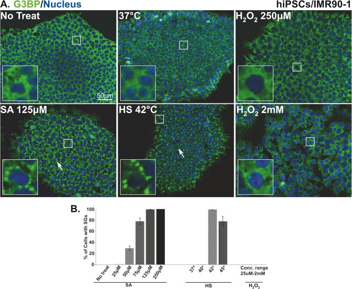
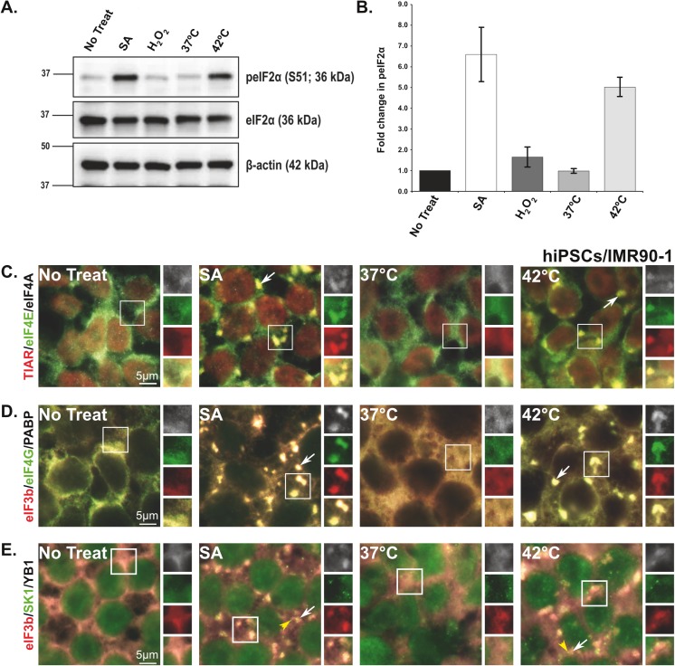
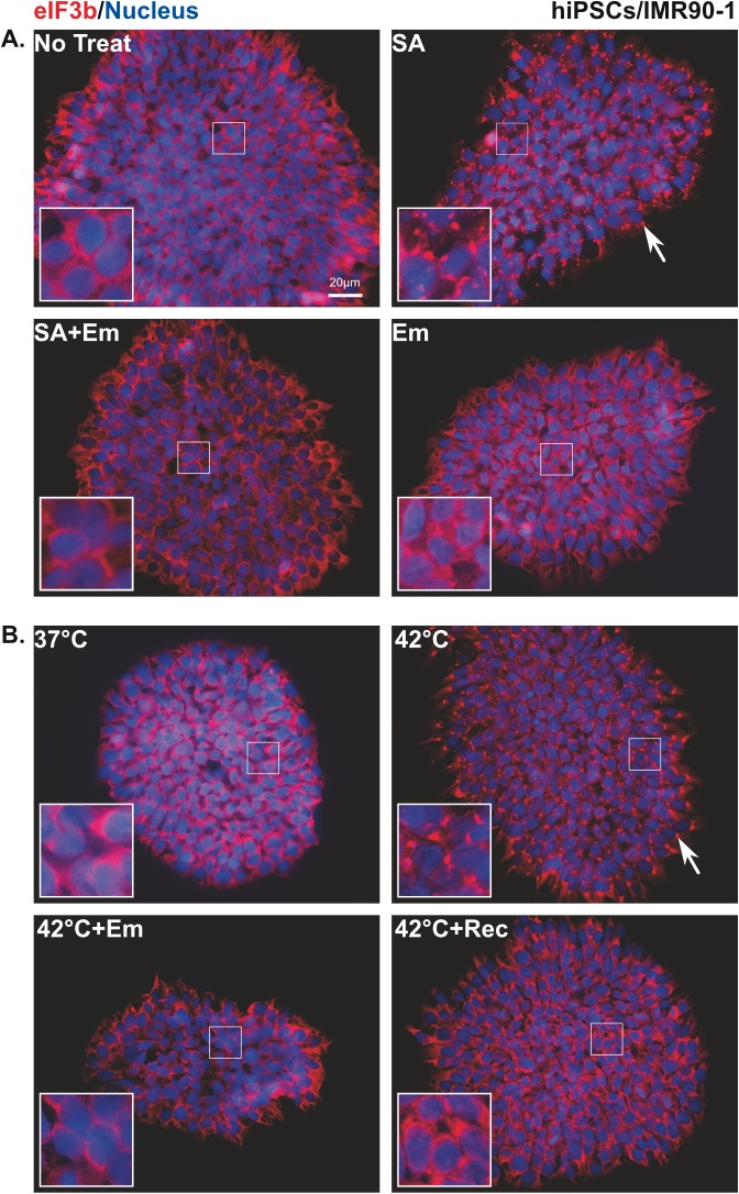
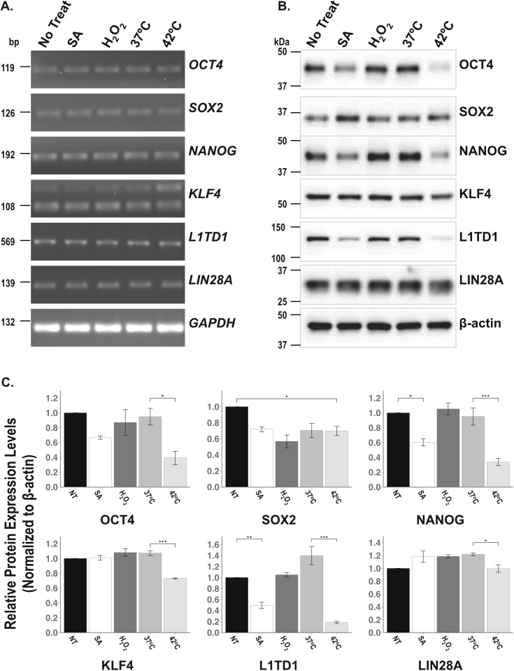
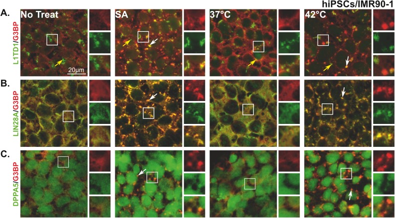
Stress Granules (SGs) are dynamic ribonucleoprotein aggregates, which have been observed in cells subjected to environmental stresses, such as oxidative stress and heat shock (HS). Although pluripotent stem cells (PSCs) are highly sensitive to oxidative stress, the role of SGs in regulating PSC self-renewal and differentiation has not been fully elucidated. Here we found that sodium arsenite (SA) and HS, but not hydrogen peroxide (H2O2), induce SG formation in human induced (hi) PSCs. Particularly, we found that these granules contain the well-known SG proteins (G3BP, TIAR, eIF4E, eIF4A, eIF3B, eIF4G, and PABP), were found in juxtaposition to processing bodies (PBs), and were disassembled after the removal of the stress. Moreover, we showed that SA and HS, but not H2O2, promote eIF2α phosphorylation in hiPSCs forming SGs. Analysis of pluripotent protein expression showed that HS significantly reduced all tested markers (OCT4, SOX2, NANOG, KLF4, L1TD1, and LIN28A), while SA selectively reduced the expression levels of NANOG and L1TD1. Finally, in addition to LIN28A and L1TD1, we identified DPPA5 (pluripotent protein marker) as a novel component of SGs. Collectively, these results provide new insights into the molecular cues of hiPSCs responses to environmental insults.


Conflict of interest statement

Competing Interests: The authors have declared that no competing interests exist.

Figures

Fig 1
Fig 1. SG assembly in human induced pluripotent stem cells is stress specific.
(A) Representative fluorescence microscopy images showing non-treated hiPSCs (No treat and 37°C), cells treated with 125μM sodium arsenite and H2O2 (250μM and 2mM) or subjected to heat shock (42°C) stained with the robust SG marker (G3BP (green)). Nucleus is stained in blue (Hoechst). Insets show magnified views of SGs. White arrows indicate SGs. Scale bar indicates 50μm. (B) Percentage of hiPSCs with G3BP positive SGs after 1h treatment with the indicated concentrations of sodium arsenite, or concentration range of H2O2 (25, 50, 100, 250, 500μM, 1mM, and 2 mM) or subjected to heat shock at the indicated temperatures. The average percentage of cells with SGs is shown. Error bars indicate the ± standard deviation from 3 independent experiments.
Fig 2
Fig 2. Sodium arsenite and heat shock treatments induce eIF2α phosphorylation and localize stress granule proteins in human induced pluripotent stem cells.
(A and B) SA and HS treatments induce eIF2α phosphorylation and SG formation in hiPSCs. (A) Representative Immuno-blots that show the levels of peIF2α (Ser51) and eIF2α in non-treated cells (No treat and 37°C) or cells treated with 125μM sodium arsenite and 250μM H2O2, or subjected to heat shock (42°C). (B) Bar graph representing the ratio of (normalized against β-actin loading controls) peIF2α (Ser51)/eIF2α for each treatment. Values are expressed as mean ± SEM from 4–5 experiments. Asterisks indicate a statistically significant change, *p<0.05, **p<0.001, and ***p<0.0001. (C-E) Representative fluorescence microscopy images showing hiPSCs treated with sodium arsenite (125μM), subjected to heat shock (42°C), or left untreated (No treat; 37°C), and stained with SG markers (C) TIAR (red)/eIF4E (green)/ eIF4A (white), (D) eIF3b (red)/eIF4G (green)/PABP (white), and (E) eIF3b (red)/YB1 (white) or PB marker (SK1, green). Insets are reproduced at the right as replicate views of SGs showing each marker separately and the merged view (yellow). Scale bar indicates 5μm.
Fig 3
Fig 3. Emetine inhibits stress granule formation in stressed human induced pluripotent stem cells.
(A) Cells treated with sodium arsenite or (B) heat shock (42°C) followed by emetine treatment (SA+Em; 42 oC+Em) or recovered from HS (42 oC+Rec) and stained with SG marker eIF3b (red). Nucleus is stained in blue (Hoechst). Insets show magnified views of SGs. White arrows indicate SGs and yellow arrowhead indicate PBs. Scale bar indicates 20μm.
Fig 4
Fig 4. Down-regulation of some pluripotent protein expression is stress specific.
(A) RT-PCR on mRNA extracted from non-treated hiPSCs (No treat and 37°C) or cells subjected to sodium arsenite, H2O2, and heat shock, to detect pluripotent genes, OCT4, SOX2, NANOG, KLF4, L1TD1, and LIN28A. GAPDH is used as a loading control. (B) Representative Immuno-blots illustrating the protein levels of OCT4, SOX2, NANOG, KLF4, L1TD1, and LIN28A. (C) Bar graphs represent the levels (normalized with β-actin loading controls) of the tested proteins. Asterisks indicate statistical significance p values; *p<0.05, **p<0.001 and ***p<0.0001. The values are expressed in mean ± SEM from 3 experiments.
Fig 5
Fig 5. Pluripotent RNA binding proteins, L1TD1, LIN28A, and DPPA5 are components of SGs.
Representative fluorescence microscopy images showing non-treated hiPSCs (No treat and 37°C) and cells treated with sodium arsenite (125μM) or subjected to heat shock (42°C) stained with the SG marker (G3BP (red)) and different pluripotent markers (A) L1TD1, (B) LIN28A, or (C) DPPA5 (green). At the right of each panel, insets show magnified views of SGs in individual and merged channels (yellow). White arrows indicate SGs and yellow arrows indicate L1TD1 aggregates. Scale bar indicates 20μm.

References

    1. Spriggs KA, Bushell M, Willis AE. Translational regulation of gene expression during conditions of cell stress. Mol Cell. 2010;40(2):228–37. doi: 10.1016/j.molcel.2010.09.028 . - DOI - PubMed
    1. Fahling M. Surviving hypoxia by modulation of mRNA translation rate. J Cell Mol Med. 2009;13(9A):2770–9. doi: 10.1111/j.1582-4934.2009.00875.x ; PubMed Central PMCID: PMCPMC4498934. - DOI - PMC - PubMed
    1. Kedersha NL, Gupta M, Li W, Miller I, Anderson P. RNA-binding proteins TIA-1 and TIAR link the phosphorylation of eIF-2 alpha to the assembly of mammalian stress granules. J Cell Biol. 1999;147(7):1431–42. ; PubMed Central PMCID: PMCPMC2174242. - PMC - PubMed
    1. Shenton D, Smirnova JB, Selley JN, Carroll K, Hubbard SJ, Pavitt GD, et al. Global translational responses to oxidative stress impact upon multiple levels of protein synthesis. J Biol Chem. 2006;281(39):29011–21. doi: 10.1074/jbc.M601545200 . - DOI - PubMed
    1. Emara MM, Fujimura K, Sciaranghella D, Ivanova V, Ivanov P, Anderson P. Hydrogen peroxide induces stress granule formation independent of eIF2alpha phosphorylation. Biochem Biophys Res Commun. 2012;423(4):763–9. doi: 10.1016/j.bbrc.2012.06.033 ; PubMed Central PMCID: PMCPMC3399031. - DOI - PMC - PubMed

MeSH terms