. 2017 Nov;38(11):5421-5439.
doi: 10.1002/hbm.23731. Epub 2017 Jul 26.

Mothers with substance addictions show reduced reward responses when viewing their own infant's face

Affiliations

Mothers with substance addictions show reduced reward responses when viewing their own infant's face

Sohye Kim et al. Hum Brain Mapp. 2017 Nov.

Abstract

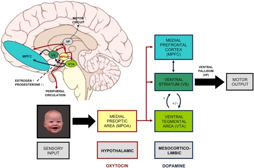
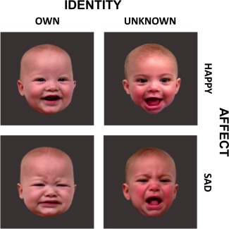
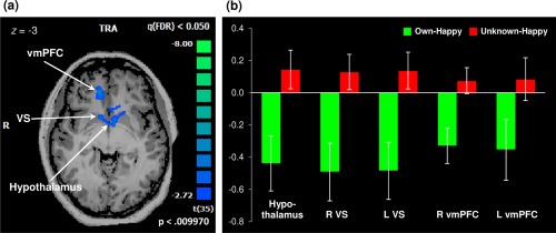
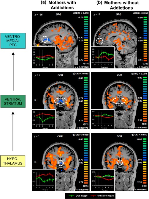
Maternal addiction constitutes a major public health problem affecting children, with high rates of abuse, neglect, and foster care placement. However, little is known about the ways in which substance addiction alters brain function related to maternal behavior. Prior studies have shown that infant face cues activate similar dopamine-associated brain reward regions to substances of abuse. Here, we report on a functional MRI study documenting that mothers with addictions demonstrate reduced activation of reward regions when shown reward-related cues of their own infants. Thirty-six mothers receiving inpatient treatment for substance addiction were scanned at 6 months postpartum, while viewing happy and sad face images of their own infant compared to those of a matched unknown infant. When viewing happy face images of their own infant, mothers with addictions showed a striking pattern of decreased activation in dopamine- and oxytocin-innervated brain regions, including the hypothalamus, ventral striatum, and ventromedial prefrontal cortex-regions in which increased activation has previously been observed in mothers without addictions. Our results are the first to demonstrate that mothers with addictions show reduced activation in key reward regions of the brain in response to their own infant's face cues. Hum Brain Mapp 38:5421-5439, 2017. © 2017 Wiley Periodicals, Inc.

Keywords: addiction; dopamine; infant; maternal; oxytocin; reward.


Figures

Figure 1
Figure 1
Hypothalamic and mesocorticolimbic regulation of maternal brain response to infant cues. These OT‐ and DA‐innervated brain regions are critical for the occurrence of maternal behavior. Brain schematic by P. J. Lynch [2006] (CC BY 2.5). [Color figure can be viewed at http://wileyonlinelibrary.com]
Figure 2
Figure 2
Infant face images used in the study. [Color figure can be viewed at http://wileyonlinelibrary.com]
Figure 3
Figure 3
Reduced maternal brain response in mothers with addictions evidenced by a single cluster of deactivation observed for the own‐happy versus unknown‐happy contrast, encompassing the hypothalamic, bilateral VS, and bilateral vmPFC regions (258 voxels, P < 0.01, FDR‐corrected q < 0.05; Panel A). Striking deactivation is observed when mothers with addictions view their own infant's smiling faces, compared with unknown infant faces (Panel B). For a post hoc examination of local peaks within the deactivated cluster, percent BOLD signal values were extracted from cubes centered at the peak voxels of each anatomical region and extending up to ± 10 voxels per dimension, unless limited by the spatial extent of activation. Peak Talairach coordinates: hypothalamus, (–2, 1, −3); R VS, (8, 10, −3); L VS, (–13, 19, 3); R vmPFC, (11, 34, 6), L vmPFC, (–16, 37, 3). Error bars represent the standard error of the mean. VS = ventral striatum; vmPFC = ventromedial prefrontal cortex; R: right; L: left. [Color figure can be viewed at http://wileyonlinelibrary.com]
Figure 4
Figure 4
Striatal activation limited to the dorsal striatum, in response to own‐happy versus unknown‐happy infant faces (P < 0.02, FDR‐corrected q < 0.05). A contrasting pattern of deactivation is seen in the ventral striatum. [Color figure can be viewed at http://wileyonlinelibrary.com]
Figure 5
Figure 5
Mothers with addictions (Panel A; N = 36) show decreased activation in the hypothalamus, VS, and vmPFC, which appear different from activations previously observed in mothers without addictions (Panel B; N = 39) in corresponding regions. Activation maps for mothers without addictions were generated from our previously published data [Strathearn and Kim, 2013] and are reprinted here for comparison. Both maps are thresholded at FDR‐corrected q < 0.05. Insets of averaged hemodynamic brain responses to own‐happy and unknown‐happy infant faces were obtained from cubes centered at the peak voxels of each anatomical region and extending up to ± 10 voxels per dimension, unless limited by the spatial extent of activation; y axis indicates % BOLD signal change shown with standard error bars; the two vertical lines represent the onset (0 sec) and offset (2 sec) times of the infant‐face‐stimulus presentation. VS = ventral striatum; vmPFC = ventromedial prefrontal cortex. [Color figure can be viewed at http://wileyonlinelibrary.com]

References

    1. Afonso VM, Sison M, Lovic V, Fleming AS (2007): Medial prefrontal cortex lesions in the female rat affect sexual and maternal behavior and their sequential organization. Behav. Neurosci 121:515–526. - PubMed
    1. Afonso VM, King S, Chatterjee D, Fleming AS (2009): Hormones that increase maternal responsiveness affect accumbal dopaminergic responses to pup‐ and food‐stimuli in the female rat. Horm Behav 56:11–23. - PubMed
    1. American Psychiatric Association (2000): Diagnostic and Statistical Manual of Mental Disorders, 4th ed., text rev. Washington, DC: American Psychiatric Association.
    1. Atzil S, Hendler T, Feldman R (2011): Specifying the neurobiological basis of human attachment: Brain, hormones, and behavior in synchronous and intrusive mothers. Neuropsychopharmacology 36:2603–2615. - PMC - PubMed
    1. Atzil S, Hendler T, Zagoory‐Sharon O, Winetraub Y, Feldman R (2012): Synchrony and specificity in the maternal and the paternal brain: Relations to oxytocin and vasopressin. J Am Acad Child Adolesc Psychiatry 51:798–811. - PubMed

Publication types

MeSH terms