

SERCA control of cell death and survival
- PMID: 28747251
- PMCID: PMC5748262
- DOI: 10.1016/j.ceca.2017.07.001
SERCA control of cell death and survival
Abstract
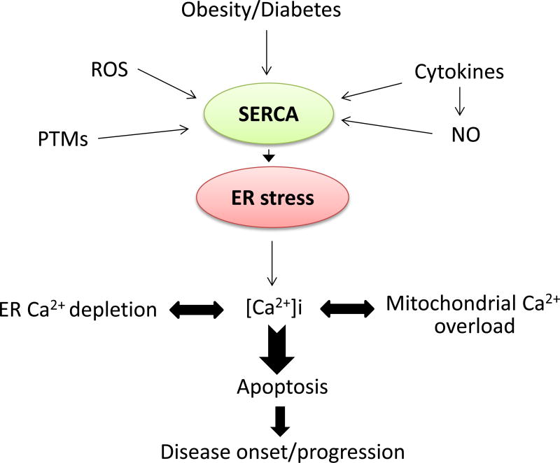
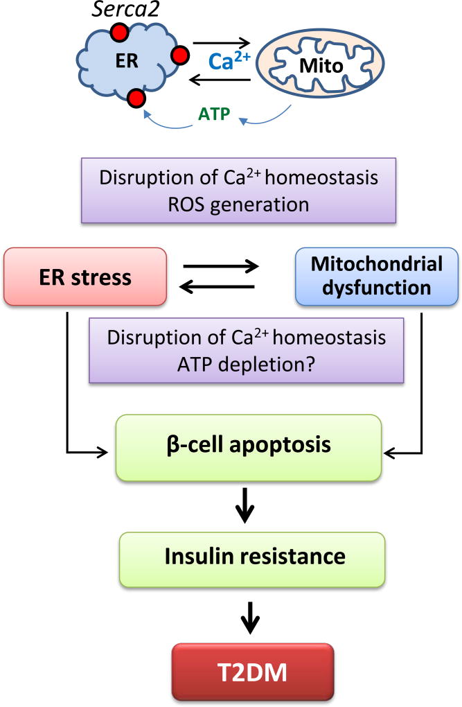
Intracellular calcium (Ca2+) is a critical coordinator of various aspects of cellular physiology. It is increasingly apparent that changes in cellular Ca2+ dynamics contribute to the regulation of normal and pathological signal transduction that controls cell growth and survival. Aberrant perturbations in Ca2+ homeostasis have been implicated in a range of pathological conditions, such as cardiovascular diseases, diabetes, tumorigenesis and steatosis hepatitis. Intracellular Ca2+ concentrations are therefore tightly regulated by a number of Ca2+ handling enzymes, proteins, channels and transporters located in the plasma membrane and in Ca2+ storage organelles, which work in concert to fine tune a temporally and spatially precise Ca2+ signal. Chief amongst them is the sarco/endoplasmic reticulum (SR/ER) Ca2+ ATPase pump (SERCA) which actively re-accumulates released Ca2+ back into the SR/ER, therefore maintaining Ca2+ homeostasis. There are at least 14 different SERCA isoforms encoded by three ATP2A1-3 genes whose expressions are species- and tissue-specific. Altered SERCA expression and activity results in cellular malignancy and induction of ER stress and ER stress-associated apoptosis. The role of SERCA misregulation in the control of apoptosis in various cell types and disease setting with prospective therapeutic implications is the focus of this review. Ca2+ is a double edge sword for both life as well as death, and current experimental evidence supports a model in which Ca2+ homeostasis and SERCA activity represent a nodal point that controls cell survival. Pharmacological or genetic targeting of this axis constitutes an incredible therapeutic potential to treat different diseases sharing similar biological disorders.
Keywords: Apoptosis; Calcium; Cancer; Cardiovascular diseases; Cell death; Diabetes; ER stress; Hepatostatosis; SERCA; SERCA isoforms; SERCA therapies.
Copyright © 2017 Elsevier Ltd. All rights reserved.
Conflict of interest statement
Figures
References
-
- Eisenberg-Lerner A, et al. Life and death partners: apoptosis, autophagy and the cross- talk between them. Cell Death Differ. 2009;16(7):966–75. - PubMed
-
- Rodriguez D, Rojas-Rivera D, Hetz C. Integrating stress signals at the endoplasmic reticulum: The BCL-2 protein family rheostat. Biochim Biophys Acta. 2011;1813(4):564–74. - PubMed
-
- Coe H, Michalak M. Calcium binding chaperones of the endoplasmic reticulum. Gen Physiol Biophys. 2009;28:F96–f103. Spec No Focus. - PubMed
-
- Groenendyk J, Michalak M. Endoplasmic reticulum quality control and apoptosis. Acta Biochim Pol. 2005;52(2):381–95. - PubMed
Publication types
MeSH terms
Substances
Grants and funding
LinkOut - more resources
Full Text Sources
Other Literature Sources
Research Materials
Miscellaneous
