. 2017 Sep 15;292(37):15352-15368.
doi: 10.1074/jbc.M116.736207. Epub 2017 Jul 26.

Thrombospondin-1 (TSP-1), a new bone morphogenetic protein-2 and -4 (BMP-2/4) antagonist identified in pituitary cells

Affiliations

Thrombospondin-1 (TSP-1), a new bone morphogenetic protein-2 and -4 (BMP-2/4) antagonist identified in pituitary cells

Céline Sallon et al. J Biol Chem. .

Abstract

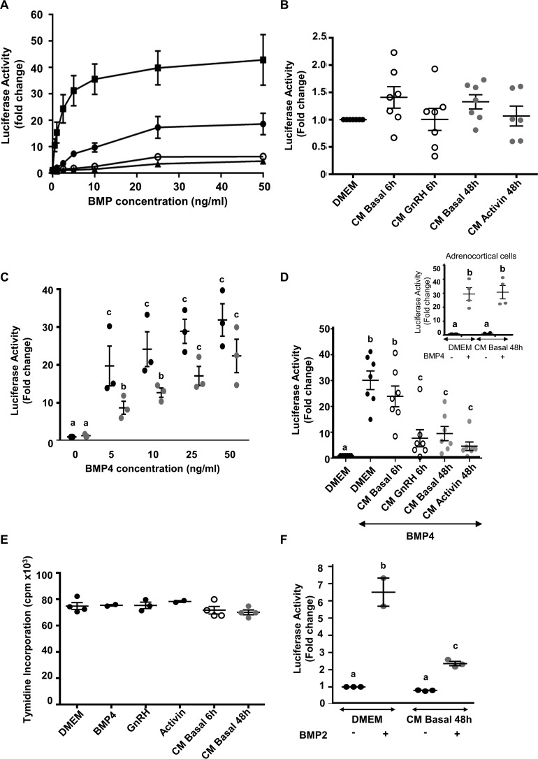
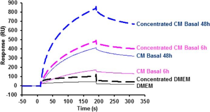
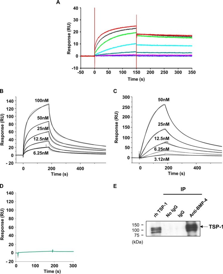
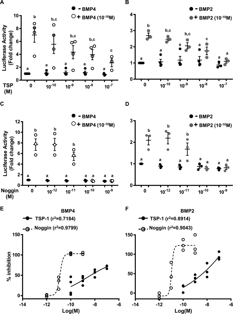
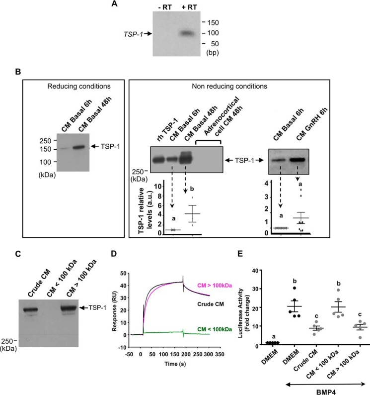
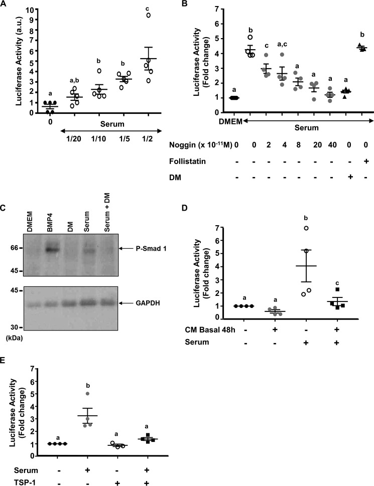
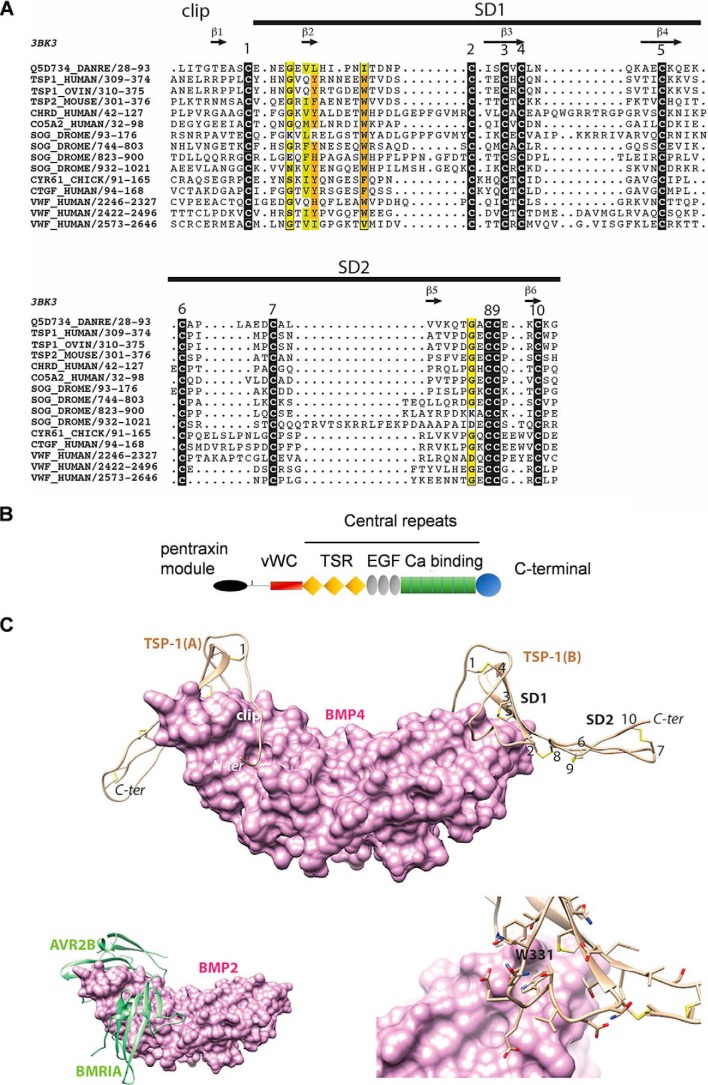
Bone morphogenetic proteins (BMPs) regulate diverse cellular responses during embryogenesis and in adulthood including cell differentiation, proliferation, and death in various tissues. In the adult pituitary, BMPs participate in the control of hormone secretion and cell proliferation, suggesting a potential endocrine/paracrine role for BMPs, but some of the mechanisms are unclear. Here, using a bioactivity test based on embryonic cells (C3H10T1/2) transfected with a BMP-responsive element, we sought to determine whether pituitary cells secrete BMPs or BMP antagonists. Interestingly, we found that pituitary-conditioned medium contains a factor that inhibits action of BMP-2 and -4. Combining surface plasmon resonance and high-resolution mass spectrometry helped pinpoint this factor as thrombospondin-1 (TSP-1). Surface plasmon resonance and co-immunoprecipitation confirmed that recombinant human TSP-1 can bind BMP-2 and -4 and antagonize their effects on C3H10T1/2 cells. Moreover, TSP-1 inhibited the action of serum BMPs. We also report that the von Willebrand type C domain of TSP-1 is likely responsible for this BMP-2/4-binding activity, an assertion based on sequence similarity that TSP-1 shares with the von Willebrand type C domain of Crossveinless 2 (CV-2), a BMP antagonist and member of the chordin family. In summary, we identified for the first time TSP-1 as a BMP-2/-4 antagonist and presented a structural basis for the physical interaction between TSP-1 and BMP-4. We propose that TSP-1 could regulate bioavailability of BMPs, either produced locally or reaching the pituitary via blood circulation. In conclusion, our findings provide new insights into the involvement of TSP-1 in the BMP-2/-4 mechanisms of action.

Keywords: C3H10T1/2 cells; bone morphogenetic protein (BMP); bone morphogenetic protein antagonist; pituitary gland; structural model; surface plasmon resonance (SPR); thrombospondin.


Conflict of interest statement

The authors declare that they have no conflicts of interest with the contents of this article

Figures

Figure 1.
Figure 1.
Effect of BMPs or pituitary CM on luciferase activity from C3H-B12 cells. A, dose-dependent induction of the BRE-Luc construct from C3H-B12 cells by BMPs. C3H-B12 cells were treated overnight with increasing concentrations of either BMP-2 (●), BMP-4 (■), BMP-6 (▴), or BMP-7 (ο) before assaying for luciferase activity as described under “Experimental Procedures.” Note that BMP-6 and BMP-7 curves are superimposed. Results are expressed as arbitrary units. Each point represents the mean ± S.E. of three experiments. B–D, effect of pituitary CM on luciferase activity from C3H-B12 cells. C3H-B12 cells were exposed overnight to DMEM or CM before assaying for luciferase activity. B, CM were from ovine pituitary cells treated or not with 10−8 m GnRH for 6 h (CM GnRH 6 h) or with 10−9 m activin for 48 h (CM activin 48 h). Values are the mean ± S.E. from seven experiments. C, pituitary media conditioned for 48 h (CM basal 48 h) were supplemented with increasing concentrations of BMP-4 before exposition to C3H-B12 cells. Values are the mean ± S.E. from three experiments. D, comparison of pituitary media conditioned for 6 h with 10−8 m GnRH (CM GnRH 6 h) or without GnRH (CM basal 6 h) or for 48 h with 10−9 m activin (CM activin 48 h) and supplemented with rh-BMP-4 (10 ng/ml) before exposition to C3H-B12 cells. Values are the mean ± S.E. from seven experiments. Inset, CM were from ovine adrenocortical cell cultures conditioned for 48 h (CM Basal 48) supplemented with BMP-4 (10 ng/ml) before exposition to C3H-B12 cells. Values are the mean ± S.E. from three experiments. E, effect of pituitary CM on C3H-B12 cell proliferation. C3H-B12 cells were exposed overnight to DMEM supplemented or not with BMP-4 (10 ng/ml), GnRH (10−8 m), or activin (10−9 m) or to pituitary CM (CM basal 6 h or CM basal 48 h) before assaying for cell proliferation. F, effect of pituitary media conditioned for 48 h (CM basal 48 h) and supplemented with BMP-2 (10 ng/ml) on luciferase activity from C3H-B12 cells. Values are the mean ± S.E. from three experiments. Bars with different letters indicate that group means are significantly different at p < 0.05.
Figure 2.
Figure 2.
Interaction between pituitary conditioned media and BMP-4 analyzed by surface plasmon resonance. BMP-4 was immobilized at intermediate density (4600 RU) on a flow cell of a CM5 sensorchip, and DMEM-BSA or 110-diluted DMEM-BSA or medium conditioned for 6 h (CM basal 6 h) or for 48 h (CM basal 48 h) diluted 110 were then injected for 180 s over the chip at 30 μl/min, and dissociation was studied for 120 s (plain lines). The sensorgrams are subtracted with the nonspecific interaction values obtained on an activated-deactivated control flow cell. Spotted lines represent aliquots of media concentrated over PEG as described under “Results” and 150-diluted before injection. The figure shows one representative experiment. Similar results were obtained with CM provided by six independent pituitary cultures.
Figure 3.
Figure 3.
Interaction between rhTSP-1 and BMP-4. A, interaction between rhTSP-1 and BMP-4 analyzed by surface plasmon resonance using BMP-4 immobilized sensorchip. To reduce TSP avidity effects, BMP-4 was immobilized at low (60 RU) density on a flow cell of a CM5 sensorchip. Increasing concentrations of rhTSP-1 (from bottom to top: 1, 2, 4, 8, 16, 32, 64 nm) were then injected for 180 s over the chip at 30 μl·min−1. Dissociation was studied for 600 s (cut here after 200 s to better see the first phase of association). The sensorgrams are double-subtracted for the nonspecific interaction and for the buffer effect. B, interaction between rhTSP-1 and BMP-4 analyzed by surface plasmon resonance using TSP-1 immobilized sensorchip. TSP-1 was immobilized at 1500 RU on a flow cell of a CM5 sensorchip. Increasing concentrations of BMP-4 from 6.25 to 100 nm were then injected for 180 s at 10 μl·min−1. C, interaction between rhTSP-1 and BMP-2 analyzed by surface plasmon resonance using TSP-1-immobilized sensorchip. TSP-1 was immobilized at 1500 RU on a flow cell of a CM5 sensorchip. Increasing concentrations of BMP-2 from 3.12 to 50 nm were then injected for 180 s at 10 μl·min−1. D, interaction between rhTSP-1 and rh-activin A analyzed by surface plasmon resonance. rhTSP-1 was immobilized on a CM5 sensorchip, and 100 nm activin A (a) or BMP-4 (b) were then injected over the chip at 10 μl/min. E, interaction between rhTSP-1 and BMP-4 analyzed by co-immunoprecipitation (IP). BMP-4 and TSP-1 were incubated together, immunoprecipitated with anti-BMP-4 antibody, and immunoblotted with anti-TSP-1 antibody (fourth lane). The second and third lanes show the result after omission of the antibody or its replacement by nonspecific IgG, respectively. Lane 1 shows the detection of rhTSP-1 (100 ng). These experiments were performed three times with similar results.
Figure 4.
Figure 4.
Antagonization of BMP-4 or BMP-2 action on luciferase activity from C3H-B12 cells. A–D, antagonization of BMP-4 or BMP-2 action by TSP-1 (A and B) or noggin (C and D). C3H-B12 cells were exposed to BMP-4 or BMP-2 (2.5 ng/ml, i.e. 10−10 m) in the presence of increasing concentrations (10−10–10−7 m) of rhTSP-1 or (10−12–10−9 m) of noggin overnight before assaying for luciferase activity. E and F, the data points, expressed as percentages of inhibition, were fitted using nonlinear regression to the Hill equation giving IC50 values of 11.4 ± 6.3, 8.5 ± 3.9, 0.02 ± 0.001, 0.02 ± 0.003 nm (A, B, C, and D, respectively) and Hill slope factors are 0.36 ± 0.1, 0.61 ± 0.13, 7.4 ± 0.6, and 6.9 ± 0.8 (A, B, C, and D, respectively). Values are the mean ± S.E. from at least three independent experiments with duplicate determinations. Bars with different letters indicate that group means are significantly different at p < 0.05.
Figure 5.
Figure 5.
Expression of pituitary TSP-1 and interaction between TSP-1-enriched CM fractions and BMP-4. A, TSP-1 mRNA expression in ovine pituitary cells. Cells were cultured for 48 h before RNA extraction. TSP-1 mRNA were analyzed by RT-PCR after 30 cycles (lane 2). PCR was performed with omission of reverse transcriptase (lane 1). B, TSP-1 protein expression in CM from pituitary cells. Cells were cultured for 6 h (CM basal 6 h) or 48 h (CM basal 48 h) or 6 h with GnRH (CM GnRH 6 h) before CM recovery and Western blot analysis in reducing or non-reducing conditions. CM from corticoadrenal cells were prepared under the same conditions. As a control, rhTSP-1 (50 ng) was loaded. Protein loading was checked by membrane staining with Ponceau S. The histograms represent the mean ± S.E. of densitometric analysis obtained with CM from six independent pituitary cultures. C, the presence of TSP-1 in high molecular mass fraction from ovine pituitary CM. Media from pituitary cells conditioned for 48 h were fractionated using 100-kDa cut-off membranes. The presence of TSP-1 in the crude CM, the filtrate (CM <100 kDa), and the retentate (CM >100 kDa) was analyzed by Western blotting. Protein loading was checked by membrane staining with Ponceau S. The figure shows a representative experiment. These experiments were performed with CM from six independent pituitary cultures. D, SPR interaction between high molecular mass fraction from pituitary CM and BMP-4. The filtrate (CM <100 kDa) 110-diluted and the retentate (CM >100 kDa) 150-diluted obtained as described above were injected for 180 s at 10 μl/min on a 60 RU immobilized BMP-4 flow cell of a CM5 sensor chip. The crude CM diluted 1/10 was injected under the same conditions. The sensorgrams are subtracted with the nonspecific interaction values obtained on the activated-deactivated control flow cell. The figure shows a representative experiment. Independent experiments were performed with CM from three different pituitary cultures. E, antagonization of BMP-4 action by the high molecular mass fraction from pituitary CM on luciferase activity from C3H-B12 cells. C3H-B12 cells were exposed to the crude CM ½-diluted, the filtrate (CM <100 kDa) ½-diluted, and the retentate (CM >100 kDa) 120-diluted in the presence of rhBMP-4 (2,5 ng/ml) overnight before assaying for luciferase activity. Values are the mean ± S.E. from four independent experiments with duplicate determinations. Bars with different letters indicate that group means are significantly different at p < 0.05.
Figure 6.
Figure 6.
BMP activity in ovine serum. A, C3H-B12 cells were exposed overnight to different dilutions of ovine serum before assaying for luciferase activity. Values are the mean ± S.E. from one representative experiment with duplicate determinations. These experiments were performed three times with similar results. C3H-B12 cells were exposed overnight to ovine serum ½-diluted supplemented with increasing doses of noggin from 0 to 4 × 10⁻¹⁰ m, 2.5 × 10⁻⁶ m dorsomorphin (DM) or 10−8 m follistatin (B) or pituitary CM (CM basal 48 h) diluted ½ (D) or 10−7 m rhTSP-1 (E) before assaying for luciferase activity. Values are the mean ± S.E. from four experiments with duplicate determinations. C, activation of SMAD1 in C3H-B12 cells by ovine serum. C3H-B12 cells were exposed for 1 h to BMP-4 (10 ng/ml) or serum ½-diluted or serum ½-diluted supplemented with dorsomorphin (2.5 × 10⁻⁶ m). Total proteins were isolated from the cells, and Western blotting was performed with phospho-SMAD1 antibody. Equal loading of the proteins was confirmed by using GAPDH antibody. Bars with different letters indicate that group means are significantly different at p < 0.05. a.u., absorbance units.
Figure 7.
Figure 7.
A, multiple sequence alignment of the VWC domain of TSP-1 with other VWC domains from the chordin family, including that of CV-2 (Q5D734_DANRE) whose 3D structure is known (PDB ID 3bk3). Black boxes indicate highly conserved cysteine residues. Observed secondary structures and disulfide bonds are reported above and below the alignment, respectively. Sequences are designated with their UniProt identifiers. B, schematic diagram of domain architecture of TSP-1 (modified from Ref. 43) vWC = vWC domain; TSR = thrombospondin type 1 domain repeats; EGF = EGF like domain repeats; Ca binding = calcium-binding type 3 repeats. C, top, model of the 3D structure of the TSP-1 VWC domain (ribbon representation) in complex with BMP-4 (surface representation). The three subdomains (clip, SD1, and SD2) are shown together with the five disulfide bridges (numbering is as depicted in Fig. 1). The model was built based on the 3D structure of CV-2 in complex with BMP-2 (PDB ID 3BK3) (34) and on the alignment shown in Fig. 1. Bottom left, experimental structure of BMP-2 bound to its high-affinity type I receptor BMPR-IA and its low-affinity type II receptor ActR-IIB (PDB ID 2H62) (34) in order to show the overlap with the VWC domain-binding sites. Bottom right, focus on the Trp-331 amino acid, fitting with a groove at the surface of BMP-4.

References

    1. Wozney J. M., Rosen V., Celeste A. J., Mitsock L. M., Whitters M. J., Kriz R. W., Hewick R. M., and Wang E. A. (1988) Novel regulators of bone formation: molecular clones and activities. Science 242, 1528–1534 - PubMed
    1. Chen D., Zhao M., and Mundy G. R. (2004) Bone morphogenetic proteins. Growth Factors. 22, 233–241 - PubMed
    1. Otsuka F. (2010) Multiple endocrine regulation by bone morphogenetic protein system. Endocr. J. 57, 3–14 - PubMed
    1. Otsuka F. (2013) Multifunctional bone morphogenetic protein system in endocrinology. Acta Med. Okayama. 67, 75–86 - PubMed
    1. Paez-Pereda M., Giacomini D., Refojo D., Nagashima A. C., Hopfner U., Grubler Y., Chervin A., Goldberg V., Goya R., Hentges S. T., Low M. J., Holsboer F., Stalla G. K., and Arzt E. (2003) Involvement of bone morphogenetic protein 4 (BMP-4) in pituitary prolactinoma pathogenesis through a SMAD/estrogen receptor crosstalk. Proc. Natl. Acad. Sci. U.S.A. 100, 1034–1039 - PMC - PubMed

MeSH terms