. 2017 Sep 15;292(37):15312-15320.
doi: 10.1074/jbc.M117.799346. Epub 2017 Jul 26.

The Alzheimer's disease-protective CD33 splice variant mediates adaptive loss of function via diversion to an intracellular pool

Affiliations

The Alzheimer's disease-protective CD33 splice variant mediates adaptive loss of function via diversion to an intracellular pool

Shoib S Siddiqui et al. J Biol Chem. .

Abstract

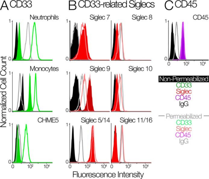
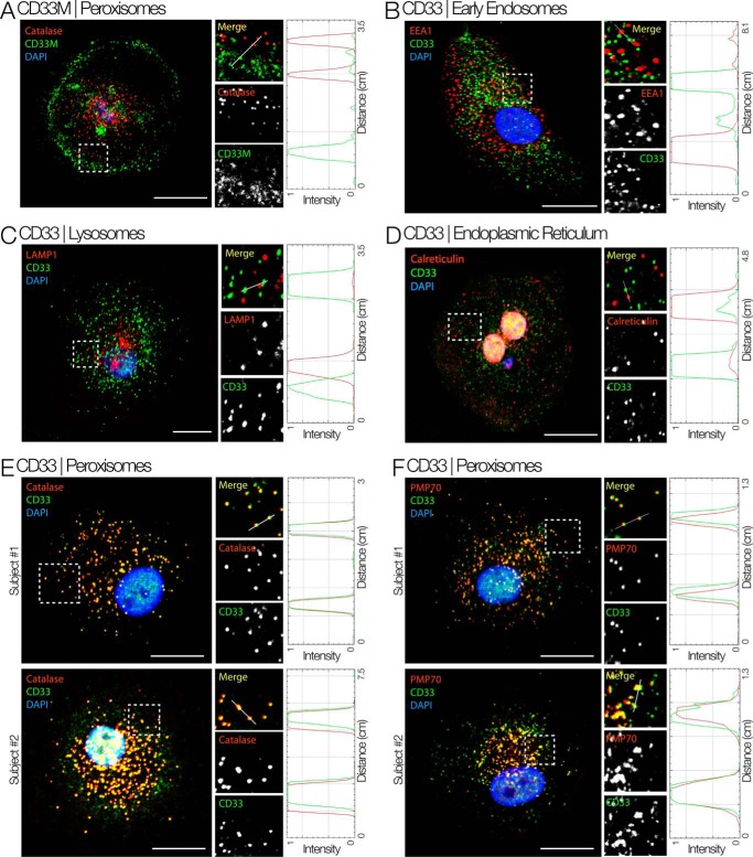
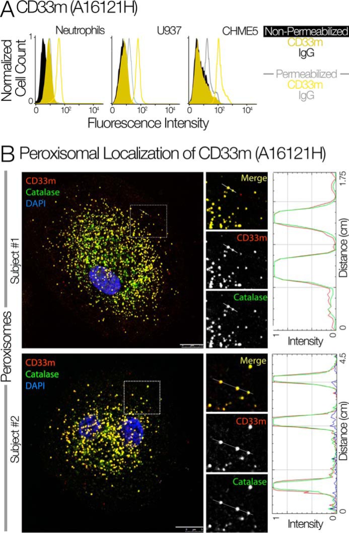
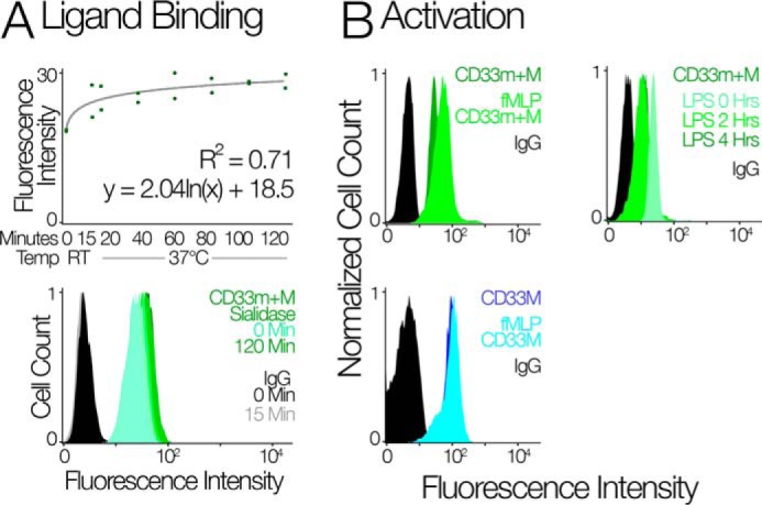
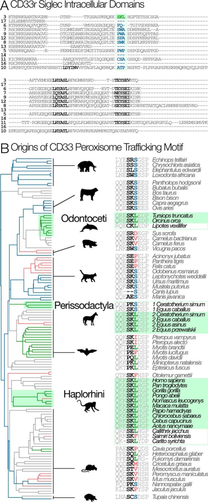
The immunomodulatory receptor Siglec-3/CD33 influences risk for late-onset Alzheimer's disease (LOAD), an apparently human-specific post-reproductive disease. CD33 generates two splice variants: a full-length CD33M transcript produced primarily by the "LOAD-risk" allele and a shorter CD33m isoform lacking the sialic acid-binding domain produced primarily from the "LOAD-protective" allele. An SNP that modulates CD33 splicing to favor CD33m is associated with enhanced microglial activity. Individuals expressing more protective isoform accumulate less brain β-amyloid and have a lower LOAD risk. How the CD33m isoform increases β-amyloid clearance remains unknown. We report that the protection by the CD33m isoform may not be conferred by what it does but, rather, from what it cannot do. Analysis of blood neutrophils and monocytes and a microglial cell line revealed that unlike CD33M, the CD33m isoform does not localize to cell surfaces; instead, it accumulates in peroxisomes. Cell stimulation and activation did not mobilize CD33m to the surface. Thus, the CD33m isoform may neither interact directly with amyloid plaques nor engage in cell-surface signaling. Rather, production and localization of CD33m in peroxisomes is a way of diminishing the amount of CD33M and enhancing β-amyloid clearance. We confirmed intracellular localization by generating a CD33m-specific monoclonal antibody. Of note, CD33 is the only Siglec with a peroxisome-targeting sequence, and this motif emerged by convergent evolution in toothed whales, the only other mammals with a prolonged post-reproductive lifespan. The CD33 allele that protects post-reproductive individuals from LOAD may have evolved by adaptive loss-of-function, an example of the less-is-more hypothesis.

Keywords: Alzheimer disease; CD33; Siglec-3; alternative splicing; intracellular trafficking; peroxisome; sialic acid.


Conflict of interest statement

The authors declare that they have no conflicts of interest with the contents of this article

Figures

Figure 1.
Figure 1.
Human innate immune cells contain an intracellular pool of CD33. A, the flow cytometry analysis of CD33 on the cell surface and intracellular in neutrophils (top panel), monocytes (middle panel), and CHME5 cell line (bottom panel). Cells were measured with their membranes intact (green-shaded distributions) and permeabilized using cytoperm/cytofix kit (green-unshaded distributions). IgG controls are in black (intact cells) and gray (permeabilized cells). B, flow cytometry analysis of other CD33rSiglecs in identically treated human neutrophils. The amount of each marker was determined by FACS using the indicated antibodies. Neutrophils were measured with their membranes intact (red-shaded distributions) and permeabilized using cytoperm/cytofix kit (red-unshaded distributions). IgG or secondary antibody controls are in black (intact cells) and gray (permeabilized cells). C, flow cytometry analysis of CD45 in identically treated human neutrophils. Cells were measured with their membranes intact (purple-shaded distributions) and permeabilized using cytoperm/cytofix kit (purple-unshaded distributions). IgG controls are in black (intact cells) and gray (permeabilized cells).
Figure 2.
Figure 2.
The intracellular pool is composed of the CD33m isoform. A, CD33 exists in two splice forms: a full length CD33M and a truncated CD33m that is lacking the V-set domain. Human alleles that predispose individuals to Alzheimer's are spliced primarily into the CD33M form, alleles that protect from Alzheimer's are spliced primarily into CD33m. B, the left panel shows the schematic regarding the reactivity of different CD33 antibodies that can partially discriminate the two forms. WM53 targets the V-set domain and labels only CD33M (blue). HIM3-4 targets the C2set domain and labels both CD33M and CD33m (green). The middle panel shows the flow cytometry analysis of CD33M and CD33m+M using the antibodies WM53 and HIM3-4 with intact and permeabilized cells. The right panel shows the quantification of mean fluorescence intensity of CD33M and CD33m+M in intact and permeabilized cells. The line shows mean fluorescence intensity. **, p < 0.01, n = 4 for CD33m+M and n = 3 for CD33M.
Figure 3.
Figure 3.
Intracellular CD33m localizes to peroxisomes. Shown is immunofluorescence staining for different cell compartments of macrophages isolated from whole blood using HIM3-4 clone. Boxed areas on left are magnified and displayed as individual channels. RGB profile plots measure the degree of co-localization of red and green pixels on vesicles along the line drawn in each magnified (Merge) panel. A, immunostaining specifically for the CD33M isoform (antibody WM53) shows that the full-length CD33M isoform is mostly located on the plasma membrane. B–F, the intracellular pool of CD33 co-localizes with peroxisome markers (Catalase and PMP70) but not with markers of early endosome (EEA1), lysosome (LAMP1), or endoplasmic reticulum (Calreticulin). Scale bar = 10 μm.
Figure 4.
Figure 4.
Confirmation of peroxisomal localization of CD33m using isoform-specific antibodies. A, the flow cytometry analysis of CD33m in neutrophils, U937, and CHME-5 cells using the newly generated CD33m antibody (Clone A16121H). B, the immunofluorescence staining of CD33m and catalase showed the peroxisomal targeting of CD33m in monocyte-derived macrophages (n = 2).
Figure 5.
Figure 5.
CD33 localization is insensitive to activating stimuli. A, the top panel shows flow cytometry analysis of CD33 on the surface of U937 cells upon sialidase treatment. The scatter plot gives the predicted mean fluorescence intensity based on least squares logarithmic regression. Two replicates were performed for each time step: 0 and 15 min at room temperature and 20, 40, 60, 80, 100, and 120 min at 37 °C. The bottom panel shows the fluorescence intensity of CD33 in U937 cells upon sialidase treatment for different time points. Sialidase treatment with AUS to remove CD33 ligands at the cell surface did not change the distribution of CD33m+M. Thus, the intracellular pool of CD33m is not due to its inability to bind sialic acid ligands. B, the top left panel shows the FACS analysis of CD33 using HIM3-4 clone on neutrophils upon stimulation with fMLP; the top right panel shows the flow cytometry analysis of CD33 on neutrophil upon stimulation with LPS. The bottom panel shows that the flow cytometry analysis of CD33M in neutrophil upon stimulation with fMLP. Treatment of neutrophils with LPS led to a decrease in cell surface CD33, and contrary to this, treatment with fMLP led to partial mobilization of CD33m to the cell surface.
Figure 6.
Figure 6.
Phylogenetic origins of the peroxisome trafficking motif. A, alignment of the intracellular region of human CD33rSiglecs (Siglec 13 is from chimpanzee). Among human CD33rSiglecs, only CD33 has a peroxisome trafficking motif (SKL: shown in green). ITIM and ITIM-like motifs are shown in black. B, the SKL motif evolved multiple times in parallel (green boxes): Haplorhine primates, Perissodactyl ungulates (in one of two CD33-like duplicates), and Odontoceti-toothed whales. Branches are colored according to the third position of the SKL motif. S is the most likely ancestral state found in clades whose common ancestors existed at the base of the tree (blue branches). L (green branches) exist in several places in the tree and are not always associated with the SKL motif. Other amino acids at this position are colored red and are represented by red branches. Unsampled mammalian clades are shown in gray.

References

    1. Macauley M. S., Crocker P. R., and Paulson J. C. (2014) Siglec-mediated regulation of immune cell function in disease. Nat. Rev. Immunol. 14, 653–666 - PMC - PubMed
    1. Crocker P. R., Paulson J. C., and Varki A. (2007) Siglecs and their roles in the immune system. Nat. Rev. Immunol. 7, 255–266 - PubMed
    1. Schwarz F., Fong J. J., and Varki A. (2015) Human-specific evolutionary changes in the biology of siglecs. Adv. Exp. Med. Biol. 842, 1–16 - PubMed
    1. Varki A., and Angata T. (2006) Siglecs: the major subfamily of I-type lectins. Glycobiology 16, 1R–27R - PubMed
    1. Walter R. B., Raden B. W., Zeng R., Häusermann P., Bernstein I. D., and Cooper J. A. (2008) ITIM-dependent endocytosis of CD33-related Siglecs: role of intracellular domain, tyrosine phosphorylation, and the tyrosine phosphatases, Shp1 and Shp2. J. Leukoc. Biol. 83, 200–211 - PubMed

Publication types

MeSH terms