. 2017 Sep 27;91(20):e00816-17.
doi: 10.1128/JVI.00816-17. Print 2017 Oct 15.

Mutations in the E2 Glycoprotein and the 3' Untranslated Region Enhance Chikungunya Virus Virulence in Mice

Affiliations

Mutations in the E2 Glycoprotein and the 3' Untranslated Region Enhance Chikungunya Virus Virulence in Mice

David W Hawman et al. J Virol. .

Abstract

Chikungunya virus (CHIKV) is a mosquito-transmitted alphavirus that causes debilitating musculoskeletal pain and inflammation and can persist for months to years after acute infection. Although studies of humans and experimentally infected animals suggest that CHIKV infection persists in musculoskeletal tissues, the mechanisms for this remain poorly understood. To evaluate this further, we isolated CHIKV from the serum of persistently infected Rag1 -/- mice at day 28. When inoculated into naive wild-type (WT) mice, this persistently circulating CHIKV strain displayed a capacity for earlier dissemination and greater pathogenicity than the parental virus. Sequence analysis revealed a nonsynonymous mutation in the E2 glycoprotein (E2 K200R) and a deletion within the 3' untranslated region (3'-UTR). The introduction of these changes into the parental virus conferred enhanced virulence in mice, although primary tropism for musculoskeletal tissues was maintained. The E2 K200R mutation was largely responsible for enhanced viral dissemination and pathogenicity, although these effects were augmented by the 3'-UTR deletion. Finally, studies with Irf3/Irf7 -/- and Ifnar1 -/- mice suggest that the E2 K200R mutation enhances viral dissemination from the site of inoculation independently of interferon regulatory factor 3 (IRF3)-, IRF7-, and IFNAR1-mediated responses. As our findings reveal viral determinants of CHIKV dissemination and pathogenicity, their further study should help to elucidate host-virus interactions that determine acute and chronic CHIKV infection.IMPORTANCE CHIKV is a globally spreading, mosquito-transmitted virus that causes debilitating acute and chronic musculoskeletal disease in humans. The viral genetic determinants that dictate the severity of acute and chronic diseases are not understood. To improve our understanding of CHIKV pathogenesis, we evaluated a CHIKV strain isolated from the serum of chronically infected immunocompromised mice. Sequence analysis of this persistent CHIKV strain identified two mutations, an amino acid change in the E2 viral attachment protein and a deletion within the 3'-UTR of the viral genome. We identified roles for these mutations in the enhancement of viral dissemination from the inoculation site and in disease severity. These data improve our understanding of the viral determinants of CHIKV pathogenesis and adaptive changes that occur during viral persistence.

Keywords: alphavirus; chikungunya virus; viral pathogenesis; virulence determinants.


Figures

FIG 1
FIG 1
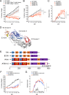
Mutations in E2 and the 3′-UTR enhance CHIKV pathogenicity in mice. (A) WT C57BL/6J mice were inoculated with 103 PFU of AF15561 (n = 5), 20 μl of clarified right ankle tissue homogenate (10 to 20 PFU) collected at day 28 p.i. from 5 individual chronically infected Rag1−/− mice (n = 5), or 20 μl of serum (10 to 20 PFU) collected from 5 individual chronically infected Rag1−/− mice (n = 5; data from individual mice are graphed separately) in the left rear footpad. The percent starting body weight was determined daily. The black arrow indicates the sample used for plaque purification of the virus. (B and C) WT C57BL/6J mice (n = 6 to 7/group) were inoculated with 10 PFU of AF15561 or plaque-purified AF15561 viral isolates derived from the serum of a chronically infected Rag1−/− mouse (AF6811P1 to AF6811P3) in the left rear footpad. The percent starting body weight (B) and disease score (C) were determined daily. Data are derived from two independent experiments. P values were determined by two-way ANOVA with Bonferroni's multiple-comparison test. **, P < 0.01; ****, P < 0.0001. (D) CHIKV E2-E1 heterodimer showing the position of E2 residue 200 (PDB accession no. 3N42) (32). E1 is shown in light gray, the E1 fusion loop is shown in magenta, E2 domain A is shown in dark blue, E2 domain B is shown in orange, and E2 domain C is shown in red. The lysine residue at E2 position 200 is shown in yellow. (E) Schematic of the 3′-UTR structures of West African (WA); East, Central, and South African (ECSA); and Asian genotype CHIKV strains, including the AF6811P2 Asian strain plaque isolated from the serum of chronically infected Rag1−/− mice. UTR sequence features were annotated according to methods described previously by Chen et al. (65). Direct repeats are labeled and illustrated by different-colored blocks. Sequence gaps in the alignment are indicated by white blocks. (F and G) WT C57BL/6J mice were inoculated with 103 PFU of AF15561 (n = 4), AF6811P2 (n = 4), or AF15561E2 K200R;ΔUTR (n = 4) in the left rear footpad. The percent starting body weight (F) and disease score (G) were determined daily. P values were determined by two-way ANOVA with Bonferroni's multiple-comparison test. ***, P < 0.001; ****, P < 0.0001.
FIG 2
FIG 2
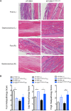
Mutations in E2 and the 3′-UTR enhance musculoskeletal tissue injury and inflammation. WT C57BL/6J mice were inoculated with 103 PFU of AF15561 or AF15561E2 K200R;ΔUTR in the left rear footpad. (A) At 7 dpi, 5-μm paraffin-embedded sections were generated from the ipsilateral and contralateral hind limbs and stained with hematoxylin and eosin. Images are representative of results for 8 mice per group. (B) Tissues in the foot, leg, and thigh from the left (L) and right (R) limbs (n = 8/group) were scored in a blind manner based on the following scale: 0 for no inflammation, 1 for >5 areas of small clusters of leukocytes, 2 for leukocytes forming larger clusters to thin tracts through tissue with multiple affected sites, 3 for clusters and tracts of leukocytes coalescing into at least one large area that displaces/replaces tissue, with or without necrosis and with or without mineralization, and 4 for leukocytes that are in aggregates that are large enough to replace >40% of normal tissue. Data are derived from results from two independent experiments. P values were determined by a Kruskal-Wallis test with Dunn's multiple-comparison test. *, P < 0.05; **, P < 0.01.
FIG 3
FIG 3
Mutations in E2 and the 3′-UTR enhance viral dissemination and tissue burdens in WT mice. WT C57BL/6 mice were inoculated with 103 PFU of AF15561 or AF15561E2 K200R;ΔUTR in the left rear footpad. At 1 dpi (n = 9 mice/group) (A) or 3 dpi (n = 5 mice/group) (B), mice were euthanized, and the amount of infectious virus in the indicated tissues was quantified by a focus-forming assay. Dashed lines indicate the limit of detection. Data are derived from results from two independent experiments. P values were determined by Mann-Whitney tests. *, P < 0.05; **, P < 0.01; ***, P < 0.001; ****, P < 0.0001.
FIG 4
FIG 4
AF15561 and AF15561E2 K200R;ΔUTR replicate similarly in vitro. Mouse embryo fibroblasts (A), differentiated C2C12 murine muscle cells (B), or C6/36 Aedes albopictus mosquito cells (C) were inoculated with AF15561 or AF15561E2 K200R;ΔUTR at an MOI of 0.1 FFU/cell (mouse embryo fibroblasts) or 0.01 FFU/cell (C2C12 and C6/36 cells). At 0 hpi (input) and 1, 6, 12, 24, 48, and 72 hpi, the amount of infectious virus present in culture supernatants was quantified by a focus-forming assay. Data are representative of results from two independent experiments. P values were determined by two-way ANOVA with Bonferroni's multiple-comparison test. **, P < 0.01; ***, P < 0.001; ****, P < 0.0001.
FIG 5
FIG 5
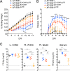
Mutations in E2 and the 3′-UTR enhance viral dissemination in Irf3−/− Irf7−/− DKO and Ifnar1−/− mice. (A and B) WT and Irf3−/− Irf7−/− DKO C57BL/6 mice were inoculated with 103 PFU of AF15561 or AF15561E2 K200R;ΔUTR in the left rear footpad. (A) At 1 dpi, the amount of infectious virus in the indicated tissues was quantified by a focus-forming assay. (B) The amount of IFN-α in serum of mock-infected (n = 3/group), AF15561-infected (n = 8-9/group), or AF15561E2 K200R;ΔUTR-infected (n = 8 to 9/group) mice was quantified by an ELISA. (C) Ifnar1−/− C57BL/6 mice were inoculated with 103 PFU of AF15561 or AF15561E2 K200R;ΔUTR in the left rear footpad. At 1 dpi, the amount of infectious virus in the indicated tissues was quantified by a focus-forming assay. Dashed lines indicate the limit of detection. Data are derived from results from two independent experiments. P values were determined by Mann-Whitney tests (A and C) or one-way ANOVA with Tukey's multiple-comparison test (B). *, P < 0.05; **, P < 0.01; ***, P < 0.001.
FIG 6
FIG 6
Both the E2 K200R mutation and the deletion in the 3′-UTR contribute to enhanced viral pathogenicity in mice. (A and B) WT C57BL/6J mice were inoculated with 103 PFU of AF15561 (n = 8), AF15561E2 K200R;ΔUTR (n = 8), AF15561E2 K200R (n = 8), or AF15561ΔUTR (n = 8) in the left rear footpad. The percent starting body weight (A) and disease score (B) were determined daily. Data are derived from results from two independent experiments. P values were determined by two-way ANOVA with Bonferroni's multiple-comparison test. **, P < 0.01; ***, P < 0.001. The P values displayed are for AF15561E2 K200R;ΔUTR versus AF15561E2 K200R. (C) WT C57BL/6J mice were inoculated with 103 PFU of AF15561 (n = 6), AF15561E2 K200R;ΔUTR (n = 6), AF15561E2 K200R (n = 6), or AF15561ΔUTR (n = 6) in the left rear footpad. At 1 dpi, mice were euthanized, and the amount of infectious virus in the indicated tissues was quantified by a focus-forming assay. Data are derived from results from two independent experiments. P values were determined by a Kruskal-Wallis test with Dunn's multiple-comparison test. *, P < 0.05; **, P < 0.01; ***, P < 0.001.

References

    1. Weaver SC, Forrester NL. 2015. Chikungunya: evolutionary history and recent epidemic spread. Antiviral Res 120:32–39. doi:10.1016/j.antiviral.2015.04.016. - DOI - PubMed
    1. Leparc-Goffart I, Nougairede A, Cassadou S, Prat C, de Lamballerie X. 2014. Chikungunya in the Americas. Lancet 383:514. doi:10.1016/S0140-6736(14)60185-9. - DOI - PubMed
    1. Pan American Health Organization. 2015. Number of reported cases of chikungunya fever in the Americas, by country or territory. Pan American Health Organization, Washington, DC: http://www.paho.org/hq/index.php?option=com_topics&view=article&id=343& Accessed 15 May 2017.
    1. Suhrbier A, Jaffar-Bandjee MC, Gasque P. 2012. Arthritogenic alphaviruses—an overview. Nat Rev Rheumatol 8:420–429. doi:10.1038/nrrheum.2012.64. - DOI - PubMed
    1. Economopoulou A, Dominguez M, Helynck B, Sissoko D, Wichmann O, Quenel P, Germonneau P, Quatresous I. 2009. Atypical Chikungunya virus infections: clinical manifestations, mortality and risk factors for severe disease during the 2005-2006 outbreak on Reunion. Epidemiol Infect 137:534–541. doi:10.1017/S0950268808001167. - DOI - PubMed