. 2017 Jul 26;7(1):6555.
doi: 10.1038/s41598-017-06848-y.

A single amino acid substitution confers B-cell clonogenic activity to the HIV-1 matrix protein p17

Affiliations

A single amino acid substitution confers B-cell clonogenic activity to the HIV-1 matrix protein p17

Cinzia Giagulli et al. Sci Rep. .

Abstract

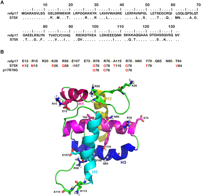
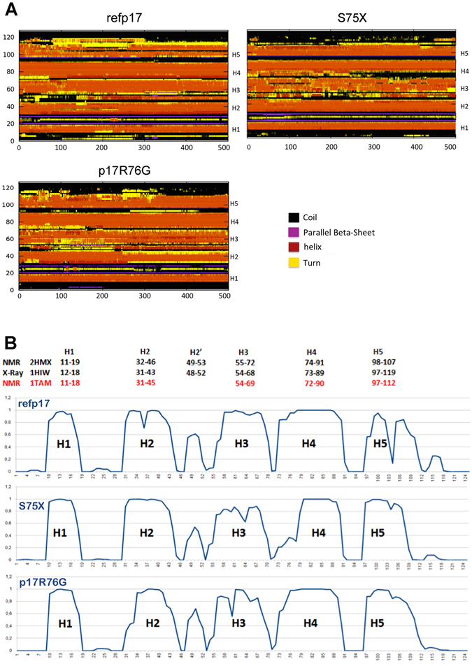
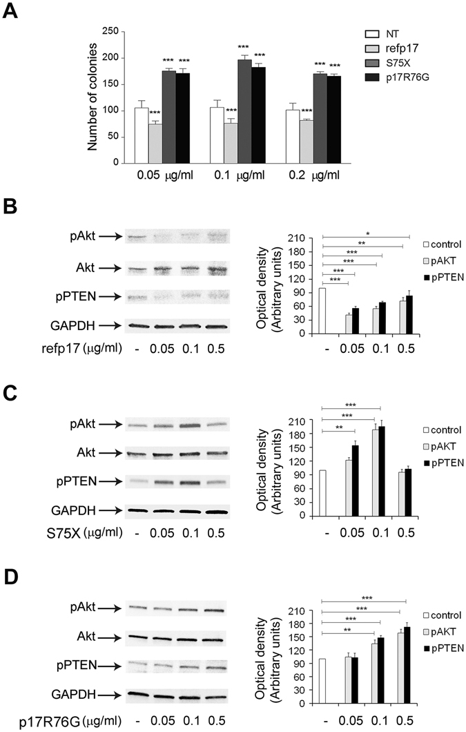
Recent data highlight the presence, in HIV-1-seropositive patients with lymphoma, of p17 variants (vp17s) endowed with B-cell clonogenicity, suggesting a role of vp17s in lymphomagenesis. We investigated the mechanisms responsible for the functional disparity on B cells between a wild-type p17 (refp17) and a vp17 named S75X. Here, we show that a single Arginine (R) to Glycine (G) mutation at position 76 in the refp17 backbone (p17R76G), as in the S75X variant, is per se sufficient to confer a B-cell clonogenic potential to the viral protein and modulate, through activation of the PTEN/PI3K/Akt signaling pathway, different molecules involved in apoptosis inhibition (CASP-9, CASP-7, DFF-45, NPM, YWHAZ, Src, PAX2, MAPK8), cell cycle promotion and cancer progression (CDK1, CDK2, CDK8, CHEK1, CHEK2, GSK-3 beta, NPM, PAK1, PP2C-alpha). Moreover, the only R to G mutation at position 76 was found to strongly impact on protein folding and oligomerization by altering the hydrogen bond network. This generates a conformational shift in the p17 R76G mutant which enables a functional epitope(s), masked in refp17, to elicit B-cell growth-promoting signals after its interaction with a still unknown receptor(s). Our findings offer new opportunities to understand the molecular mechanisms accounting for the B-cell growth-promoting activity of vp17s.


Conflict of interest statement

The authors declare that they have no competing interests.

Figures

Figure 1
Figure 1
Sequence and hydrogen bond network of refp17 and S75X. (A) Sequences are represented by the single-letter amino acid code. The amino acid sequence (aa 1–132) of the matrix protein clade B isolate BH10 p17 (UniProtKB P04585) was adopted as reference (refp17) for this analysis. Each amino acid residue of S75X not differing from refp17 sequence is represented by a dot. (B) At the top of the Figure, in the first row, the residues involved in the hydrogen bond network of p17 are shown and below the residues mutated in the variants S75X and p17R76G are indicated. The residues involved in hydrogen bond network are shown as sticks.
Figure 2
Figure 2
Evolution of refp17, S75X and p17R76G protein structure by molecular dynamics simulations. (A) Time evolution of the secondary structural elements along the molecular dynamics simulation generated by DSSP. The X-axis represents the molecular dynamics trajectory time (in ns), while the residue numbers are shown on the Y-axis. (B) At the top of the Figure are indicated the residues involved in forming the helix structures of refp17 and determined experimentally by NMR (PDB code: 2HMX and 1TAM) and X-Ray (PDB code: 1HIW). In the graph the propensity for each amino acid residue to assume helix structure calculated during simulations of refp17 and its variants.
Figure 3
Figure 3
Structure and stability of refp17, S75X and p17R76G. (A) CD spectra of refp17, S75X, and p17R76G collected at room temperature at 2.5 μM in 10 mM phosphate buffer, pH 7.4. (B) Thermal denaturation of refp17, S75X, and p17R76G at 10 μM in PBS, pH 7.4, as monitored at 222 nm by CD spectroscopy. The experimental data were normalized according to a two-state protein denaturation model. Note that the thermal denaturation of p17 is irreversible due to protein aggregation. (C) Measurement of the size of folded refp17, S75X and p17R76G viral matrix proteins by the technique of dynamic light scattering at room temperature. (D) NMR spectra of refp17 (black), S75X (green), and p17R76G (red) showing their amide-amide NOEs.
Figure 4
Figure 4
Effect of refp17, S75X and p17R76G on B-cell activity. (A) Raji were plated in twelve-well plates and, after four days, medium was replaced by fresh medium with the indicated concentration of refp17, S75X and p17R76G. Cells not treated (NT) were used as negative control. The cell growth was analyzed by using MTT. Data represent the average number of colonies ± SD from three independent experiments performed in triplicate. The statistical significance between control and treated cultures was calculated using one-way ANOVA performed separately for each concentration of p17 variants and Bonferroni’s post-test was used to compare data; ***P < 0.001. (BD) Cells were treated for 5 min with 0.05, 0.1, 0.5 μg/ml of refp17 (B), S75X (C) and p17R76G (D). Untreated cells were used as control. Western blot analysis of Raji lysates shows that refp17 inhibits the activation of Akt and maintains PTEN in an active state (B), as shown by the respective phosphorylation state at any concentration tested, verified by densitometric analysis and plotting of the pAkt/Akt and pPTEN/GAPDH. On the contrary, either S75X or p17R76G induce the activation of Akt and maintains PTEN in an inactive state (C,D), as shown by the increased phosphorylation, verified by densitometric analysis and plotting of the pAkt/Akt and pPTEN/GAPDH. In the left panel blots from one representative experiment of three with similar results are shown. In the right panels, values reported for phosphorylation of Akt and PTEN are the mean ± SD of three independent experiments. Statistical analysis was performed by one-way ANOVA and the Bonferroni’s post-test was used to compare data; *P < 0.05; **P < 0.01; ***P < 0.001.
Figure 5
Figure 5
Representation of the putative signaling pathways involved in B-cell clonogenicity induced by interaction of S75X/p17R76G with p17R(s). Stimulation of B cells with the clonogenic p17 proteins, S75X and p17R76G, induces the activation of several molecules involved in promoting cell survival, cell cycle progression and in inhibiting apoptosis, and STRING database and literature data mining were used to identify known and experimentally verified interactions. The several kinases involved in the pathway are indicated by orange diamonds, the other proteins are represented by light red ellipses. PI3K: phosphatidylinositol-3-kinase; Akt: Protein kinase B;PP2C-alpha: Protein phosphatase 1A; MAPK8: Mitogen-activated protein kinase 8; YWHAZ: 14-3-3 protein zeta/delta; CHEK2: Checkpoint kinase 2; CDK1: Cyclin-dependent kinase 1; CDK2: Cyclin-dependent kinase 2; NPM: Nucleophosmin; DFF-45: DNA fragmentation factor subunit alpha; CASP-9: caspase-9; CASP-7: caspase-7; PAK1: Serine/threonine-protein kinase PAK 1; PAX2: Paired box protein Pax-2.
Figure 6
Figure 6
Representation of KEGG pathways significantly regulated by S75X and p17R76G. The proteins modulated by p17s were used as input for the Cytoscape ClueGO functional analysis tool to cluster the proteins according to the KEGG pathways database. The proteins are represented by yellow circles. The pathways are represented by red circles and the circle size is directly proportional to statistical relevance (3 * 10−3 > p < 8 * 10−6). An edge is present when the protein is involved in the pathway.

References

    1. Shiels MS, et al. Cancer burden in the HIV-infected population in the United States. J. Natl. Cancer Inst. 2011;103:753–762. doi: 10.1093/jnci/djr076. - DOI - PMC - PubMed
    1. Simard EP, Pfeiffer RM, Engels EA. Cumulative incidence of cancer among individuals with acquired immunodeficiency syndrome in the United States. Cancer. 2011;117:1089–1096. doi: 10.1002/cncr.25547. - DOI - PMC - PubMed
    1. Carbone A, et al. Diagnosis and management of lymphomas and other cancers in HIV-infected patients. Nat. Rev. Clin. Oncol. 2014;11:223–238. doi: 10.1038/nrclinonc.2014.31. - DOI - PubMed
    1. Vandenhende MA, et al. Cancer-Related Causes of Death among HIV-Infected Patients in France in 2010: Evolution since 2000. PLoS One. 2015;10:e0129550. doi: 10.1371/journal.pone.0129550. - DOI - PMC - PubMed
    1. Epeldegui M, Vendrame E, Martínez-Maza O. HIV-associated immune dysfunction and viral infection: role in the pathogenesis of AIDS-related lymphoma. Immunol. Res. 2010;48:72–83. doi: 10.1007/s12026-010-8168-8. - DOI - PMC - PubMed

Publication types

MeSH terms

Substances