. 2016 Oct 3;2(2):vew027.
doi: 10.1093/ve/vew027. eCollection 2016 Jul.

Unbiased whole-genome deep sequencing of human and porcine stool samples reveals circulation of multiple groups of rotaviruses and a putative zoonotic infection

Affiliations

Unbiased whole-genome deep sequencing of human and porcine stool samples reveals circulation of multiple groups of rotaviruses and a putative zoonotic infection

My V T Phan et al. Virus Evol. .

Abstract

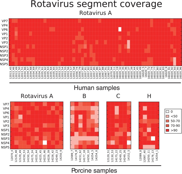
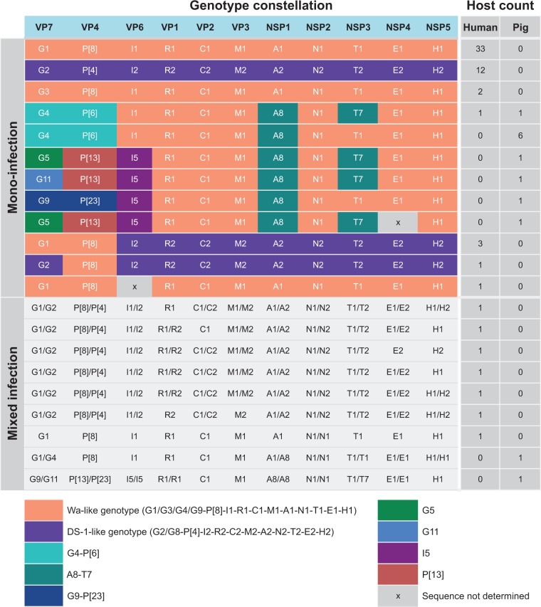
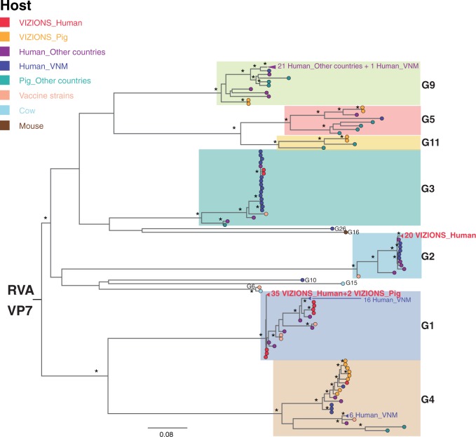
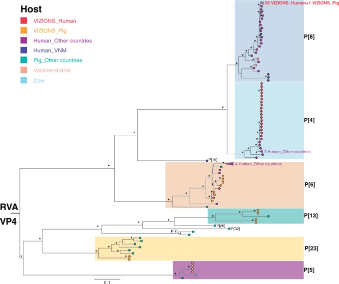
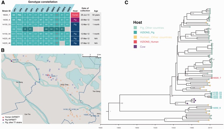
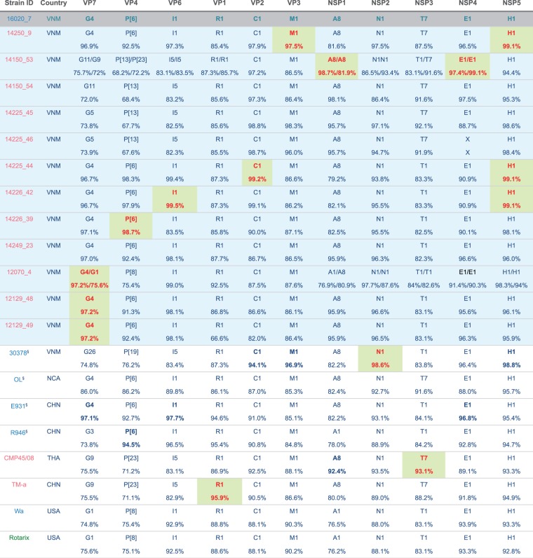
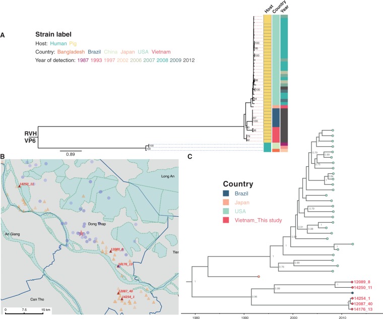
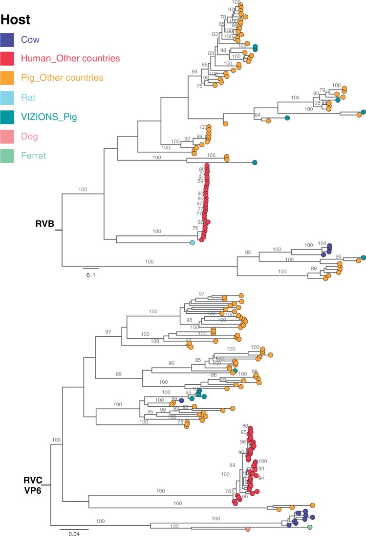
Coordinated and synchronous surveillance for zoonotic viruses in both human clinical cases and animal reservoirs provides an opportunity to identify interspecies virus movement. Rotavirus (RV) is an important cause of viral gastroenteritis in humans and animals. In this study, we document the RV diversity within co-located humans and animals sampled from the Mekong delta region of Vietnam using a primer-independent, agnostic, deep sequencing approach. A total of 296 stool samples (146 from diarrhoeal human patients and 150 from pigs living in the same geographical region) were directly sequenced, generating the genomic sequences of sixty human rotaviruses (all group A) and thirty-one porcine rotaviruses (thirteen group A, seven group B, six group C, and five group H). Phylogenetic analyses showed the co-circulation of multiple distinct RV group A (RVA) genotypes/strains, many of which were divergent from the strain components of licensed RVA vaccines, as well as considerable virus diversity in pigs including full genomes of rotaviruses in groups B, C, and H, none of which have been previously reported in Vietnam. Furthermore, the detection of an atypical RVA genotype constellation (G4-P[6]-I1-R1-C1-M1-A8-N1-T7-E1-H1) in a human patient and a pig from the same region provides some evidence for a zoonotic event.

Keywords: deep sequencing; rotavirus; virus surveillance; whole genomes; zoonotic infection.


Figures

Figure 1.
Figure 1.
Heat map of RV sequence length coverage by segment detected in all samples. The sequence length coverage for each segment of all assembled rotaviruses by deep sequencing was calculated and expressed as [(length of assembled contig in nt)/(full-length of that segment in nt)]×100. Colour code for %genome coverage is indicated in figure key ranging from low (pale orange) to high (dark red). The value of 0 indicates that contig sequence for that segment was not identified or did not pass the stringent quality control criteria including % reads mapped and contig length (see Materials and Methods). All RVA sequences detected in human samples were shown in the first panel, with each column representing a sample and each row showing each RV segment. Similarly, porcine RV samples were shown, each panel representing RVA, RVB, RVC, and RVH sequences with the segment names given vertically and sample IDs horizontally.
Figure 2.
Figure 2.
Genotype constellations of assembled RVA genomes. The genotypes of all assembled sequences were determined according to the guidelines of RV Classification Working Group, representing the genotype of VP7-VP4-VP6-VP1-VP2-VP3-NSP1-NSP2-NSP3-NSP4-NSP5. For mono-infection, each row represents one genotype constellation with the colour block used to illustrate different genotype patterns, such as common human types of Wa-like (orange) and DS-1-like (purple), and other less common genotypes shown in other colour blocks as indicated. The number of each of the genotype constellation identified in samples from human and pigs were given in the column Host count for Human and Pigs, respectively. For mixed infection where two distinct contigs were assembled for at least one segment, the genotypes of both contigs were given and indicated the host where strains were identified.
Figure 3.
Figure 3.
ML phylogenetic trees inferred from the assembled nucleotide sequences for RVA VP7 and VP4 genes. (A) ML tree of VP7 gene showed genetic relationships between sequences from this study and additional sequences of corresponding segments from GenBank. Strains were coloured according to the host species from which the strain was identified, and “VIZIONS” in the annotation refers to strains identified from this study. Tree is mid-point rooted for the purpose of clarity and bootstrap values of  ≥75 percent are shown as asterisks. All horizontal branch lengths are drawn to the scale of nucleotide substitutions per site. (B) ML tree of VP4 gene showed genetic relationships between sequences from this study and additional sequences of corresponding segments from GenBank. The pattern of tree visualization is consistent with VP7 tree, see description of Fig. 3A for more information.
Figure 3.
Figure 3.
ML phylogenetic trees inferred from the assembled nucleotide sequences for RVA VP7 and VP4 genes. (A) ML tree of VP7 gene showed genetic relationships between sequences from this study and additional sequences of corresponding segments from GenBank. Strains were coloured according to the host species from which the strain was identified, and “VIZIONS” in the annotation refers to strains identified from this study. Tree is mid-point rooted for the purpose of clarity and bootstrap values of  ≥75 percent are shown as asterisks. All horizontal branch lengths are drawn to the scale of nucleotide substitutions per site. (B) ML tree of VP4 gene showed genetic relationships between sequences from this study and additional sequences of corresponding segments from GenBank. The pattern of tree visualization is consistent with VP7 tree, see description of Fig. 3A for more information.
Figure 4.
Figure 4.
The analysis of RVA zoonotic strain G4P[6]T7 in the study. (A) The genotype constellation of the case in investigation 16020_7 and other porcine RVA strains with the NSP3 T7 genotype. Details on dates of collection and age of host are given. The colour code in the host column is consistent with colour illustration of corresponding case in the map (panel B) in this figure. (B) The geographical location of the human case’s residency and pig farms that raised the pigs infected with RVA NSP3 T7 strains overlaid on the total sampling area (as shown in Supplementary Fig. S1). The colouring of the human case and pig farms is consistent with colour code presented in the column “Host” in panel of this figure. The red star indicates the Dong Thap Provincial Hospital where diarrhoeal patients were admitted. The map scale bar is shown in the units of geometric km. See Supplementary Fig. S1 for more information. (C) The time-resolved phylogenetic tree of RVA NSP3 T7 genotype sequences comparing local versus global sequences. Reference sequences were retrieved from GenBank [N = 69, excluded the duplicate sequence for TM-a strain (JX290174) and BP1901 (KF835960) which is 15 aa shorter than the complete ORF]. Strains were coloured according to the host species from which the strain was identified, and “VIZIONS” in the annotation refers to strains identified from this study. Sequences identified from this study were highlighted in red (human case 16020_7) or in turquoise (porcine sequences). The first T7 sequence identified (in a cow in 1973) was indicated in purple. Posterior probabilities of internal nodes with values ≥0.75 are shown and the scale axis indicates time in year of strain identification.
Figure 5.
Figure 5.
Genotype constellations of RVA 16020_7 compared with representative human and animal RVA of known genotypes. Segments are bold in red and shaded in green to indicate the segments with highest nucleotide sequence similarities to that of strain 16020_7. The strain was coloured according the host from which strain was identified, blue indicates human host, pink for pigs and green for vaccine component. All porcine samples from this study were shaded in light blue and the human case of interest (16020_7) was shaded in grey. §Human strains were previously shown to have porcine origin. Country of isolation abbreviation, VNM, Vietnam; NCA, Nicaragua; CHN, China; THA, Thailand; USA, United States of America.
Figure 6.
Figure 6.
The analysis of RVH VP6 gene segment. (A) ML phylogenetic tree of RVH VP6 sequences. The RVH VP6 sequences in this study (N = 5) were compared with available full-length RVH VP6 sequences retrieved from GenBank (N = 39). Tree is mid-point rooted for the purpose of clarity and only bootstrap values of ≥ 75 percent are shown. All horizontal branch lengths are drawn to the scale of nucleotide substitutions per site in the tree. Strains were colour coded according to the host associated with the strain, the country where the strains were identified and the year of strain detection. (B) The geographical locations of pig farms that raised the pigs infected with RVH identified in this study. The farms were illustrated as red triangles with strain ID given, overlaid on the overall sampling area as shown in Supplementary Fig. S1. The red star indicates the Dong Thap Provincial Hospital. The map scale bar is shown in the units of geometric km. Refer to Supplementary Fig. S1 for more information on the background and provincial features colouring. (C) Time-resolved phylogenetic tree of porcine RVH VP6 sequences comparing local versus global sequences. Strains were coloured by the country where strains were identified. Porcine sequences identified from this study were highlighted in red, with strain ID given to link with geographical locations on map in panel B. Strains from Brazil were coloured in dark blue; orange indicates the Japanese porcine strain, and light green refers to the porcine strain from the USA. Posterior probabilities of internal nodes with values ≥0.75 are shown and the scale axis indicates time in year of strain identification.
Figure 7.
Figure 7.
ML phylogenetic trees inferred from the assembled nucleotide sequences for VP6 gene of RVB and RVC. ML trees of RVB and RVC VP6 segments showed genetic relationships between assembled sequences from this study and full-length reference sequences of corresponding segments retrieved from GenBank. Trees are mid-point rooted for the purpose of clarity and only bootstrap values of ≥ 75 percent are shown. Scale bars are in the unit of nucleotide substitutions per site. Strains were coloured according to the host species that the sequences were identified from, and “VIZIONS” in the annotation refers to strains identified from this study.

Similar articles

Cited by

References

    1. Alam M. M., et al. (2013) ‘The First Identification of Rotavirus B from Children and Adults with Acute Diarrhoea in Kathmandu, Nepal’, Tropical Medicine and Health, 41: 129–34. - PMC - PubMed
    1. Amimo J. O., Vlasova A. N., Saif L. J. (2013) ‘Prevalence and Genetic Heterogeneity of Porcine Group C Rotaviruses in Nursing and Weaned Piglets in Ohio, USA and Identification of a Potential New VP4 Genotype’, Veterinary Microbiology, 164: 27–38. - PMC - PubMed
    1. Anderson E. J., Weber S. G. (2004) ‘Rotavirus Infection in Adults’, Lancet Infectious Diseases, 4: 91–9. - PMC - PubMed
    1. Anh D. D., et al. (2006) ‘The Burden of Rotavirus Diarrhea in Khanh Hoa Province, Vietnam: Baseline Assessment for a Rotavirus Vaccine Trial’, Pediatrics Infectious Disease Journal, 25: 37–40. - PubMed
    1. Armah G. E., et al. (2010) ‘Efficacy of Pentavalent Rotavirus Vaccine Against Severe Rotavirus Gastroenteritis in Infants in Developing Countries in Sub-Saharan Africa: A Randomised, Double-Blind, Placebo-Controlled Trial’, Lancet, 376: 606–14. - PubMed