Review
. 2017 Sep;8(5):513-527.
doi: 10.1016/j.jare.2017.04.005. Epub 2017 Apr 28.

Epidemiology, pathophysiology, and management of uric acid urolithiasis: A narrative review

Affiliations
Review

Epidemiology, pathophysiology, and management of uric acid urolithiasis: A narrative review

A Abou-Elela. J Adv Res. 2017 Sep.

Abstract

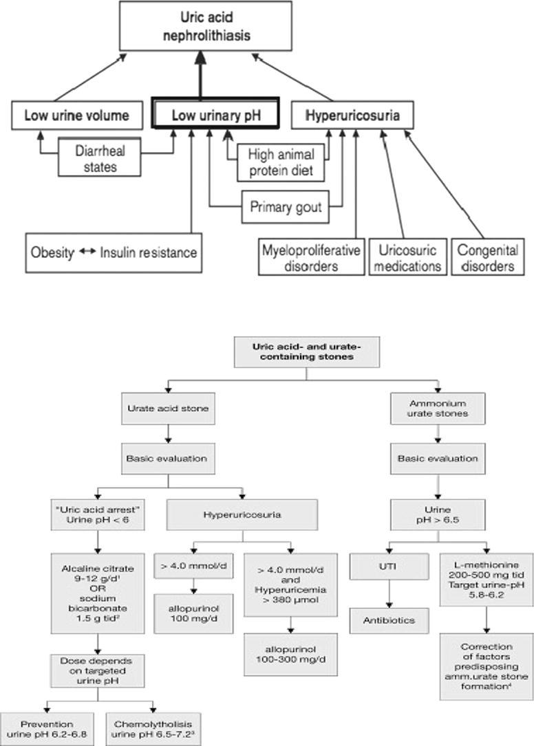
An in-depth comprehension of the epidemiology as well as pathophysiology of uric acid urolithiasis is important for the identification, treatment, and prophylaxis of calculi in these patients. Persistently low urinary pH, hyperuricosuria, and low urinary volume are the most important factors in pathogenesis of uric acid urolithiasis. Other various causes of calculus formation comprises of chronic diarrhea, renal hyperuricosuria, insulin resistance, primary gout, extra purine in the diet, neoplastic syndromes, and congenital hyperuricemia. Non-contrast-enhanced computed tomography is the radiologic modality of choice for early assessment of patients with renal colic. Excluding situations where there is acute obstruction, rising blood chemistry, severe infection, or unresolved pain, the initial management ought to be medical dissolution by oral chemolysis since this method has proved to be effective in most of the cases.

Keywords: Calculi; Chemolysis; Nephrolithiasis; Uric acid; Uric acid stones; Urinary stones; Urolithiasis; pH dissolution.


Figures

None
Graphical abstract
Fig. 1
Fig. 1
(A and B) Gross appearance and endoscopic view of uric acid stones.
Fig. 2
Fig. 2
Radiographic imaging of uric acid stone. (A) KUB demonstrating lack of radiopaque stone. (B) IVP showing filling defect in left renal pelvis. (C) CT scan with corresponding stone demonstrated .
Fig. 3
Fig. 3
Urology system – X-ray and ultrasound stone localization for radiolucent (uric acid calculi).
Fig. 4
Fig. 4
(A–C) Fluoroscopy image showing steps of dilatation & disappearance of waist during dilatation using balloon dilatation set.
Fig. 5
Fig. 5
(A–C) An X-ray showing a branching (stag-horn) stone, extracted with open surgery by anatrophic nephrolithotomy where the renal pedicle is clamped and kidney is cooled.
Fig. 6
Fig. 6
Treatment algorithm for renal calculi .
Fig. 7
Fig. 7
(A and B) Ureteroscopy endoscopic view showing stone LASER intracorporeal lithotripsy and extraction with a Dormia basket.
Fig. 8
Fig. 8
Recommended treatment options (if indicated for active stone removal) .

References

    1. Ngo Tin C., Assimos Dean G. Uric acid nephrolithiasis: recent progress and future directions. Rev Urol. 2007;9(1):17–27. - PMC - PubMed
    1. Moe O.W. Uric acid nephrolithiasis: proton titration of an essential molecule? Curr Opin Nephrol Hypertens. 2006;15:366–373. - PubMed
    1. Gianfrancesco F., Esposito T., Ombra M.N., Forabosco P., Maninchedda G., Fattorini M. Identification of a novel gene and a common variant associated with uric acid nephrolithiasis in a Sardinian genetic isolate. Am J Hum Genet. 2003;72:1479–1491. - PMC - PubMed
    1. Daudon M, Frochot V. Crystalluria. Clinical chemistry and laboratory medicine 2015;53(Suppl 2):s1479–87. - PubMed
    1. Gul Z., Monga M. Medical and dietary therapy for kidney calculus prevention. Kor J Urol. 2014;55(12):775–779. - PMC - PubMed

LinkOut - more resources