Review
. 2018 Jan;75(2):275-290.
doi: 10.1007/s00018-017-2602-1. Epub 2017 Jul 26.

Mechanisms of protein homeostasis (proteostasis) maintain stem cell identity in mammalian pluripotent stem cells

Affiliations
Review

Mechanisms of protein homeostasis (proteostasis) maintain stem cell identity in mammalian pluripotent stem cells

Alireza Noormohammadi et al. Cell Mol Life Sci. 2018 Jan.

Abstract

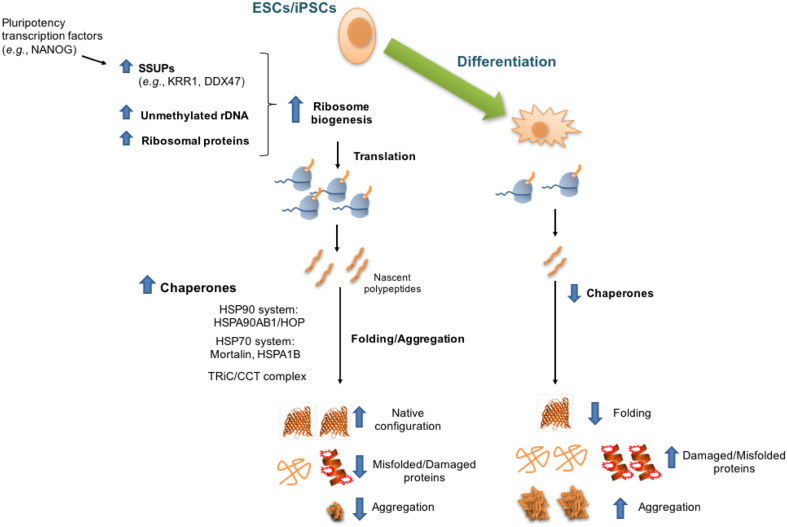
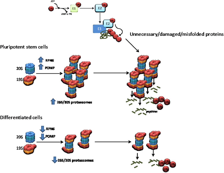
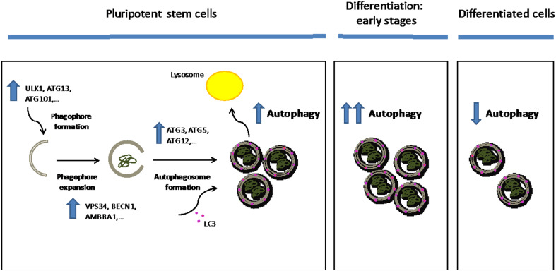
Protein homeostasis, or proteostasis, is essential for cell function, development, and organismal viability. The composition of the proteome is adjusted to the specific requirements of a particular cell type and status. Moreover, multiple metabolic and environmental conditions challenge the integrity of the proteome. To maintain the quality of the proteome, the proteostasis network monitors proteins from their synthesis through their degradation. Whereas somatic stem cells lose their ability to maintain proteostasis with age, immortal pluripotent stem cells exhibit a stringent proteostasis network associated with their biological function and intrinsic characteristics. Moreover, growing evidence indicates that enhanced proteostasis mechanisms play a central role in immortality and cell fate decisions of pluripotent stem cells. Here, we will review new insights into the melding fields of proteostasis and pluripotency and their implications for the understanding of organismal development and survival.

Keywords: Autophagy; Chaperones; Differentiation; Pluripotency; Proteasome; Proteostasis; Stress responses.


Conflict of interest statement

The authors declare that they have no conflict of interest.

Figures

Fig. 1
Fig. 1
Intrinsic regulation of proteostasis-related anabolic processes of pluripotent stem cells. Scheme showing specific components of protein translation and folding nodes, which are differentially expressed in pluripotent stem cells compared with their differentiated counterparts. Pluripotent stem cells exhibit up-regulated global levels of protein synthesis rates, a process modulated by enhanced expression of several subunits of the small subunit processome (SSUP), unmethylated state of rDNA promoter, and translation of ribosome subunits. To assist the folding of high amounts of newly synthesized proteins, pluripotent stem cell function requires increased expression of chaperone core machinery genes. In addition, the intrinsic chaperome network of pluripotent stem cells maintains the proper folded state of proteins from their synthesis through their degradation, resulting in decreased accumulation of damaged/misfolded proteins and aggregates. Although differentiated cells exhibit decreased translational rates, a concomitant down-regulation in the chaperone/folding system diminishes their ability to refold damaged/misfolded proteins and restrain protein aggregation
Fig. 2
Fig. 2
Intrinsic regulatory mechanisms of 26S/30S proteasome activity in pluripotent stem cells compared with their differentiated counterparts. Proteasome-mediated degradation starts with the sequential attachment of ubiquitin molecules to the target substrate (e.g., regulatory, unnecessary, damaged, and misfolded proteins), a process mediated by a three-step cascade mechanism. First, ubiquitin is activated in an ATP-dependent manner by the ubiquitin-activating enzyme (E1). The activated ubiquitin is transferred to ubiquitin-conjugating enzymes (E2s) via formation of E2-ubiquitin thioester structure. Then, E3 ligases catalyze the attachment of ubiquitin to their specific target by binding both the E2-ubiquitin thioester structure and the substrate protein. The same sequential mechanism links additional molecules to the primary ubiquitin through internal ubiquitin lysines, forming a polyubiquitin chain. E3 ligases provide specificity to the proteasomal-degradation process and over 600 E3 ligases have been identified in humans so far. After the polyubiquitination cascade process, target proteins are recognized and degraded by the 26S/30S proteasome. Active 26S/30S proteasomes are formed by the interaction between the 20S catalytic core and the 19S regulatory particle. Pluripotent stem cells exhibit increased levels of RPN6 and POMP resulting in increased assembly and activity of the 26S/30S proteasome. On the contrary, the levels of RPN6 and POMP are down-regulated in differentiated cells resulting in decreased assembly of 26S/30S proteasomes
Fig. 3
Fig. 3
Autophagic flux in pluripotency and differentiation. The ULK complex (ULK1, ATG13, FIP200, and ATG101) regulates the formation of the double membrane structure known as the phagophore. Then, the VPS34–BECN1 complex (formed by VPS34, BECN1, AMBRA1, and ATG14L) mediates the expansion of the phagophore. Once the cytoplasmic fraction is engulfed into the phagophore, the membrane elongates until it closes forming the autophagosome, a process regulated by the ATG12–ATG5–ATG16L1 complex, which enhances the conjugation of cytosolic LC3 (LC3I) to phosphatidylethanolamine. LC3–phosphatidylethanolamine conjugate (LC3II) is recruited to the membrane and then LC3II-containing autophagosomes are trafficked to the lysosome for their degradation. Pluripotent stem cells exhibit a higher basal autophagic flux compared to terminally differentiated cells such as fibroblast and neurons. These up-regulated levels of autophagic flux are sustained by increased expression of core genes involved in different steps of autophagy. Moreover, autophagy–lysosome activity is further up-regulated during early differentiation steps. Once the cells are differentiated, they exhibit decreased autophagic flux compared with undifferentiated pluripotent stem cells

References

    1. Balchin D, Hayer-Hartl M, Hartl FU. In vivo aspects of protein folding and quality control. Science. 2016;353(6294):aac4354. doi: 10.1126/science.aac4354. - DOI - PubMed
    1. Hartl FU. Cellular homeostasis and aging. Annu Rev Biochem. 2016;85:1–4. doi: 10.1146/annurev-biochem-011116-110806. - DOI - PubMed
    1. Balch WE, et al. Adapting proteostasis for disease intervention. Science. 2008;319(5865):916–919. doi: 10.1126/science.1141448. - DOI - PubMed
    1. Powers ET, et al. Biological and chemical approaches to diseases of proteostasis deficiency. Annu Rev Biochem. 2009;78:959–991. doi: 10.1146/annurev.biochem.052308.114844. - DOI - PubMed
    1. Labbadia J, Morimoto RI. The biology of proteostasis in aging and disease. Annu Rev Biochem. 2015;84:435–464. doi: 10.1146/annurev-biochem-060614-033955. - DOI - PMC - PubMed

Publication types