Review
. 2017 Oct;35(10):2105-2114.
doi: 10.1002/stem.2667. Epub 2017 Jul 26.

Concise Review: Stem Cells for Corneal Wound Healing

Affiliations
Review

Concise Review: Stem Cells for Corneal Wound Healing

Mehrnoosh Saghizadeh et al. Stem Cells. 2017 Oct.

Abstract

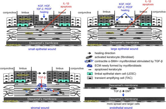
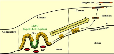
Corneal wound healing is a complex process that occurs in response to various injuries and commonly used refractive surgery. It is a significant clinical problem, which may lead to serious complications due to either incomplete (epithelial) or excessive (stromal) healing. Epithelial stem cells clearly play a role in this process, whereas the contribution of stromal and endothelial progenitors is less well studied. The available evidence on stem cell participation in corneal wound healing is reviewed, together with the data on the use of corneal and non-corneal stem cells to facilitate this process in diseased or postsurgical conditions. Important aspects of corneal stem cell generation from alternative cell sources, including pluripotent stem cells, for possible transplantation upon corneal injuries or in disease conditions are also presented. Stem Cells 2017;35:2105-2114.

Keywords: Cell transplantation; Corneal endothelium; Corneal epithelium; Gene therapy; Keratocyte; Pluripotent stem cell; Stem cell; Wound healing.


Figures

Figure 1
Figure 1
Schematic representation of main events during corneal epithelial, stromal, and endothelial wound healing. Top left, healing of small epithelial wound under the influence of several growth factors entails participation of central cells only. Keratocytes under the wound die by apoptosis mediated by epithelium‐derived interleukin‐1β. Top right, healing of large epithelial wound under the influence of several growth factors entails participation of both limbal epithelial stem cells and their progeny (transient amplifying cells), as well as of central cells. Bottom left, healing of a stromal wound entails activation of keratocytes to form fibroblasts that are transformed to motile myofibroblasts under the influence of transforming growth factor (TGF)‐β. Myofibroblasts positive for α‐smooth muscle actin contract the wound, and also produce and remodel the extracellular matrix in the wound bed. Burns are also associated with stromal neovascularization (not shown). Bottom right, healing of endothelial wound entails epithelial–mesenchymal transformation (EMT) and cell migration under the influence of TGF‐β. Wound closure is accompanied by increased spreading and enlargement of endothelial cells that undergo the process opposite to EMT, that is, mesenchymal–epithelial transformation. Abbreviations: ECM, extracellular matrix; EMT, epithelial–mesenchymal transformation; HGF, hepatocyte growth factor; IGF‐2, insulin‐like growth factor‐2; IL, interleukin; KGF, keratinocyte growth factor; PEDF, pigment epithelium‐derived factor; TGF, transforming growth factor; α‐SMA, α‐smooth muscle actin.
Figure 2
Figure 2
Corneal epithelial cell maintenance by limbal epithelial stem cell (LESC). LESC (expressing K14, K15, and p63α, and potentially other markers) residing in the basal epithelium of the palisades of Vogt, divide (X) and differentiate into transient amplifying cells while they migrate centripetally (Y), first horizontally along the basement membrane then diagonally through the epithelial tiers, before reaching the superficial epithelium in the central cornea as terminally differentiated cells that are sloughed from the ocular surface (Z). According to Thoft and Friend, X + Y = Z. Reproduced with permission from 8. Abbreviations: BV, blood vessel; LESC, limbal epithelial stem cell; PoV, palisades of Vogt; TAC, transient amplifying cell; TDC, terminally differentiated cell.
Figure 3
Figure 3
Clonal expansion and participation of limbal epithelial stem cell in corneal epithelial homeostasis and wound healing. Left, 6‐week old transgenic mice were injected intraperetoneally with tamoxifen over 3 consecutive days. Mice were monitored long‐term by intravital microscopy, as described previously. Colored patches were observed in the peripheral cornea at 3 weeks (W) post‐tamoxifen (A, arrows), which developed into discrete stripes (B, C; 8 and 11 weeks post‐tamoxifen, respectively) migrating toward the central cornea intersecting the apex by 18 weeks (D). Notably, the intraocular lens autofluoresces and the eyelid skin recombined within 1 week of tamoxifen treatment. Scale bar = 400 μm. Reproduced with permission from 8. Right, expansion and migration of K14+ fluorescent clones in Confetti transgenic mice during wound healing. Chemical burns to the corneal surface were achieved by topical application of dimethyl sulfoxide (DMSO) combined with tamoxifen induction. (D): Repeated DMSO application for 3 successive days caused a “severe” wound to the cornea. One week following the last DMSO treatment, multiple wide streaks of fluorescent cells were observed; (D′) is a magnification of (D). Limbal–corneal border is annotated by a dashed line. Scale bar = 500 μm (D); = 75 μm (D′). Reproduced with permission from 17. Abbreviations: CFP, cyan fluorescent protein; GFP, green fluorescent protein; RFP, red fluorescent protein; YFP, yellow fluorescent protein.
Figure 4
Figure 4
Re‐epithelialization of the corneal surface by human amniotic membrane (HAM)‐grown limbal epithelial stem cell‐enriched limbal cultures. Left, low magnification showing HAM with attached limbal cells placed on top of human organ cultured cornea that has been de‐epithelialized by mild NaOH treatment. HAM is secured to the corneal surface by derma + flex gel adhesive (formulated medical cyanoacrylate from Chemence Medical, Alpharetta, GA). Scale bar = 100 μm. Right, high magnification showing limbal cells that have migrated from HAM and repopulated the corneal surface. Scale bar = 40 μm. Abbreviation: HAM, human amniotic membrane.
Figure 5
Figure 5
Debridement‐wounded mouse corneas were treated with fibrin gel only (no cells) or with 50,000 limbal biopsy‐derived stromal cells in fibrin gel. After 4 weeks of healing, histological sections (epithelium oriented up) were stained for fibrotic markers decorin, biglycan, tenascin C, fibronectin, and hyaluronan binding protein. Images are representative of sections from three corneas for each condition. Note lack of fibrotic proteins from the stem cell‐treated corneas that are now similar to the untreated ones. Reproduced with permission from 76. Abbreviation: LBSC, limbal biopsy‐derived stromal cell.

Similar articles

Cited by

References

    1. Ljubimov AV, Saghizadeh M. Progress in corneal wound healing. Prog Retin Eye Res 2015;49:17–45. - PMC - PubMed
    1. Davanger M, Evensen A. Role of the pericorneal papillary structure in renewal of corneal epithelium. Nature 1971;229:560–561. - PubMed
    1. Tseng SC. Regulation and clinical implications of corneal epithelial stem cells. Mol Biol Rep 1996;23:47–58. - PubMed
    1. Dua HS, Shanmuganathan VA, Powell‐Richards AO et al. Limbal epithelial crypts: A novel anatomical structure and a putative limbal stem cell niche. Br J Ophthalmol 2005;89:529–532. - PMC - PubMed
    1. Shortt AJ, Secker GA, Munro PM et al. Characterization of the limbal epithelial stem cell niche: Novel imaging techniques permit in vivo observation and targeted biopsy of limbal epithelial stem cells. Stem Cells 2007;25:1402–1409. - PubMed

Publication types