Review
. 2017 Jul 27:3:17048.
doi: 10.1038/nrdp.2017.48.

Oesophageal cancer

Affiliations
Review

Oesophageal cancer

Elizabeth C Smyth et al. Nat Rev Dis Primers. .

Abstract

Oesophageal cancer is the sixth most common cause of cancer-related death worldwide and is therefore a major global health challenge. The two major subtypes of oesophageal cancer are oesophageal squamous cell carcinoma (OSCC) and oesophageal adenocarcinoma (OAC), which are epidemiologically and biologically distinct. OSCC accounts for 90% of all cases of oesophageal cancer globally and is highly prevalent in the East, East Africa and South America. OAC is more common in developed countries than in developing countries. Preneoplastic lesions are identifiable for both OSCC and OAC; these are frequently amenable to endoscopic ablative therapies. Most patients with oesophageal cancer require extensive treatment, including chemotherapy, chemoradiotherapy and/or surgical resection. Patients with advanced or metastatic oesophageal cancer are treated with palliative chemotherapy; those who are human epidermal growth factor receptor 2 (HER2)-positive may also benefit from trastuzumab treatment. Immuno-oncology therapies have also shown promising early results in OSCC and OAC. In this Primer, we review state-of-the-art knowledge on the biology and treatment of oesophageal cancer, including screening, endoscopic ablative therapies and emerging molecular targets, and we discuss best practices in chemotherapy, chemoradiotherapy, surgery and the maintenance of patient quality of life.


Conflict of interest statement

Conflict of interest

ES declares honararia for advisory role from Five Prime Therapeutics and Bristol-Meyer Squibb. DC declares institutional research funding from Amgen, AstraZeneca, Bayer, Celgene, MedImmune, Merck Serono, Merrimack, and Sanofi. FL has received research support from GlaxoSmithKline and Fresenius Biotech; lecture and advisory honoraria from Amgen, Biontech, Bristol-Myers Squibb, Eli Lilly, Ganymed, Merck-Serono, MSD, Nordic and Roche; travel support from Amgen, Bayer, Roche and Taiho. MAS declares honararia for an adivory role from Lilly and institutional research Funding from Genentech and Sanofi. JL, PL, RF declare no conflicts.

Figures

Figure 1
Figure 1
Annual incidence rate of OAC and OSCC globally in men. Panel a demonstrates highest rates for OAC in Western industrialised nations including USA, Canada, Australia and European Union, and lower levels in less developed areas including Africa and China. Panel B demonstrates high levels of OSCC in China, in so called “oesophageal cancer belt” across extending to Iran, and also in East Africa.
Figure 2
Figure 2
Pathogenesis of oesophageal squamous cell carcinoma and adenocarcinoma. The oesophageal mucosal is exposed to repeated insult (tobacco, alcohol, hot liquids, reflux containing acid or bile) which results in changes to the squamous oesophageal mucosa. Molecular changes also accumulate ultimately leading to a malignant phenotype. In OSCC squamous hyperplasia precedes low and high grade squamous dysplasia which then develops into invasive cancer. In OAC, a metaplastic epithelium containing intestinal metaplasia is transformed through low and high grade dysplasia to invasive cancer.
Figure 3
Figure 3
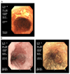
A. Endoscopy image demonstrating low grade dysplasia in squamous mucosal lesion at 12 o’clock highlighted using Lugol’s solution. B. Endoscopy image demonstrating intramucosal squamous lesion in a patient with achalasia. .
Figure 4
Figure 4
Eight edition TNM categories. T is categorized as Tis: high-grade dysplasia (HGD). T1 is cancer that invades the lamina propria, muscularis mucosae, or submucosa and is subcategorized into T1a (cancer that invades the lamina propria or muscularis mucosae) and T1b (cancer that invades the submucosa); T2 is cancer that invades the muscularis propria; T3 is cancer that invades the adventitia; T4 is cancer that invades the local structures and is subcategorized as T4a (cancer that invades adjacent structures such as the pleura, pericardium, azygos vein, diaphragm, or peritoneum) and T4b (cancer that invades the major adjacent structures, such as the aorta, vertebral body, or trachea). N is categorized as N0 (no regional lymph node metastasis), N1 (regional lymph node metastases involving one to two nodes), N2 (regional lymph node metastases involving three to six nodes), and N3 (regional lymph node metastases involving seven or more nodes). M is categorized as M0 (no distant metastasis) and M1 (distant metastasis). Reproduced from Thomas et al. Cancer of the Esophagus and Esophagogastric Junction: An Eighth Edition Staging Primer. Journal of Thoracic Oncology, vol 12, n 1: 36-42 (2016).
Figure 5
Figure 5
Suggested algorithm for management of localised oesophageal cancer
Figure 6
Figure 6
These pictures illustrate a minimally invasive oesophagectomy for oesophageal cancer. Picture A shows the port placement of the abdominal part of the procedure. Picture B shows how the greater omentum has been mobilised and is pulled towards the abdominal wall. Picture C shows the arteries that join and constitute the celiac trunk following lymphadenectomy (the stomach is pushed against the abdominal wall). Picture D shows the port placement of the thoracic part of the procedure with the patients lying in his abdomen. Picture E shows how the oesophagus is mobilised and dissected from adjacent tissue (the right lunch is collapsed). Picture F shows the chest after the oesophagus has been resected, visualising the airways, pericardium and stapled proximal oesophagus and the proximal part of the gastric tube
Figure 7
Figure 7
Pathways of interest and available drugs in development in oesophageal cancer.

References

    1. Lin Y, et al. Epidemiology of esophageal cancer in Japan and China. Journal of epidemiology. 2013;23:233–242. - PMC - PubMed
    1. Cook MB, Chow WH, Devesa SS. Oesophageal cancer incidence in the United States by race, sex, and histologic type, 1977-2005. British journal of cancer. 2009;101:855–859. - PMC - PubMed
    1. Edgren G, Adami H-O, Weiderpass Vainio E, Nyrén O. A global assessment of the oesophageal adenocarcinoma epidemic. Gut. 2012 - PubMed
    1. The Cancer Genome Atlas Research, N. Integrated genomic characterization of oesophageal carcinoma. Nature. 2017;541:169–175. - PMC - PubMed
    1. Fitzgerald RC, et al. British Society of Gastroenterology guidelines on the diagnosis and management of Barrett's oesophagus. Gut. 2014;63:7–42. - PubMed

MeSH terms

Supplementary concepts