. 2017 Jul 27;13(7):e1005686.
doi: 10.1371/journal.pcbi.1005686. eCollection 2017 Jul.

Upregulation of an inward rectifying K+ channel can rescue slow Ca2+ oscillations in K(ATP) channel deficient pancreatic islets

Affiliations

Upregulation of an inward rectifying K+ channel can rescue slow Ca2+ oscillations in K(ATP) channel deficient pancreatic islets

Vehpi Yildirim et al. PLoS Comput Biol. .

Abstract

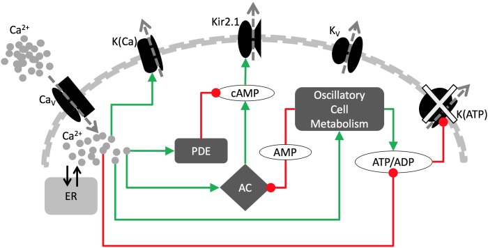
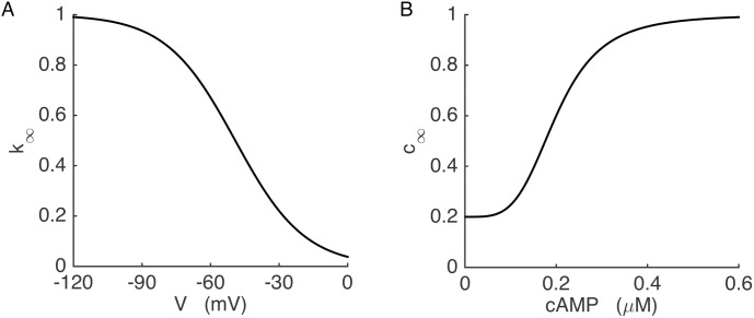
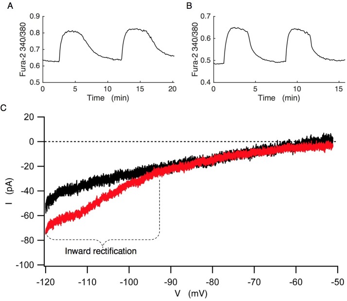
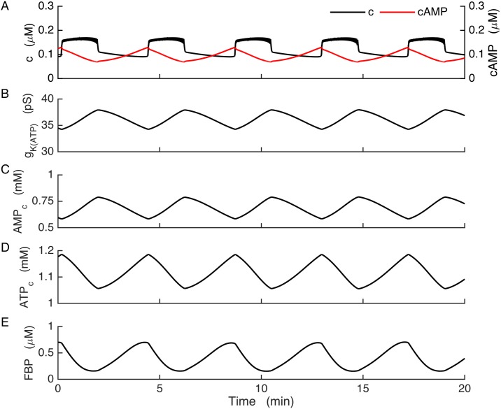
Plasma insulin oscillations are known to have physiological importance in the regulation of blood glucose. In insulin-secreting β-cells of pancreatic islets, K(ATP) channels play a key role in regulating glucose-dependent insulin secretion. In addition, they convey oscillations in cellular metabolism to the membrane by sensing adenine nucleotides, and are thus instrumental in mediating pulsatile insulin secretion. Blocking K(ATP) channels pharmacologically depolarizes the β-cell plasma membrane and terminates islet oscillations. Surprisingly, when K(ATP) channels are genetically knocked out, oscillations in islet activity persist, and relatively normal blood glucose levels are maintained. Compensation must therefore occur to overcome the loss of K(ATP) channels in K(ATP) knockout mice. In a companion study, we demonstrated a substantial increase in Kir2.1 protein occurs in β-cells lacking K(ATP) because of SUR1 deletion. In this report, we demonstrate that β-cells of SUR1 null islets have an upregulated inward rectifying K+ current that helps to compensate for the loss of K(ATP) channels. This current is likely due to the increased expression of Kir2.1 channels. We used mathematical modeling to determine whether an ionic current having the biophysical characteristics of Kir2.1 is capable of rescuing oscillations that are similar in period to those of wild-type islets. By experimentally testing a key model prediction we suggest that Kir2.1 current upregulation is a likely mechanism for rescuing the oscillations seen in islets from mice deficient in K(ATP) channels.


Conflict of interest statement

The authors have declared that no competing interests exist.

Figures

Fig 1
Fig 1. The key components of the model.
Green arrows are for stimulatory and red circles are for inhibitory pathways. In the wild-type cells, bursting is paced by metabolic oscillations acting on K(ATP) channels. In the KO cells, genetic disruption of K(ATP) channels leads to increased Kir2.1 current, which now drives bursting.
Fig 2
Fig 2. The Kir2.1 channel conductance depends on voltage and the cAMP concentration.
(A) Voltage-dependent blockade of the Kir2.1 current. (B) cAMP-dependent activation of the Kir2.1 current.
Fig 3
Fig 3
Fura-2 Ca2+ measurements from wild-type (A) and SUR1-/- islets (B) at 11 mM glucose. The change in Ca2+ is expressed as the Fura-2 340/380 ratio. (C) Comparison of I-V curves from wild-type (black) and SUR1-/- (red) β-cells. The wild-type recording is representative of n = 6 islets isolated from 4 mice. The SUR1-/- recording is representative of n = 8 islets isolated from 5 mice. The SUR1-/- islets exhibited significant inward rectification at more negative potentials compared to cells from wild-type islets.
Fig 4
Fig 4. Bursting in wild-type model cells.
Slow glycolytic oscillations drive bursting through actions on the K(ATP) current. (A) cAMP declines at the start of each Ca2+ plateau. (B) K(ATP) channel conductance. (C-D) Adenine nucleotide concentrations in the cytosol. (E) Slow glycolytic oscillations are reflected in the FBP time course.
Fig 5
Fig 5. Bursting in the model KO cells, where K(ATP) current is replaced with Kir2.1 current.
Glycolytic oscillations drive bursting through a cAMP-dependent pathway. (A) Ca2+ and cAMP concentrations oscillate in anti-phase. (B) Conductance of the Kir2.1 current, time averaged over a window of 6 s to remove fast variations and highlight the cAMP-dependent slow dynamics. (C) AMPc oscillations contribute to the production of cAMP oscillations. (D) ATPc oscillates due to oscillations in glycolysis. (E) FBP is the product of the PFK enzyme that is responsible for glycolytic oscillations. For this simulation, the glucokinase reaction rate was increased from 0.09 μM/ms to 0.14 μM/ms and kFBP was increased from 0.8 to 0.95.
Fig 6
Fig 6. In the model KO cells, bursting is driven by the Kir2.1 current, which is regulated by voltage and the cAMP concentration.
(A) Mean V and c during a burst. Voltage is averaged over each spike. (B) The cAMP and cytosolic AMP concentrations. (C) Dynamics of the Kir2.1 channel activation (c) and inactivation (k). (D) Kir2.1 conductance during a burst.
Fig 7
Fig 7. Fura-2 Ca2+ measurements of islets in 11 mM glucose and, as indicated, 50 μM of the membrane permeable 8-Br-cAMP.
(A) Ca2+ oscillations in wild-type islets persist with little or no change upon application of 8-Br-cAMP. Representative of 10 islets. (B) Ca2+ oscillations in SUR1-/- islets are terminated by 8-Br-cAMP, and Ca2+ is at a low level. Representative of 9 islets.
Fig 8
Fig 8. Glycolytic oscillations drive bursting in the model KO cell.
(A) c (black) oscillates reflecting bursting electrical activity, while AMPc oscillates (blue) reflecting glycolytic oscillations. (B) Bifurcation diagram of the fast subsystem, with c as bifurcation parameter. HB = Hopf bifurcation, SN = saddle-node bifurcation, SNIC = saddle-node on invariant circle bifurcation. Solid and dashed curves represent stable and unstable steady states, respectively, while bold solid and bold dashed curves represent stable and unstable limit cycles, respectively. (C) The burst trajectory projected onto the c-V plane. (D) Fast/slow analysis of bursting, with the burst trajectory (red) and c curve superimposed onto the fast-subsystem bifurcation diagram. The c curve is shown for AMPc at its minimum (dashed magenta) and maximum (dashed green) during a burst.
Fig 9
Fig 9. The model KO cell can produce bursting with upregulation of a constant-conductance (leak) K+ current and a K(Ca) conductance: gleak = 32.5 pS, gK(Ca) = 90 pS.
(A) Negative feedback of c (black) on the membrane potential and slow cer (blue) oscillations drive bursting. (B) The fast-subsystem bifurcation diagram exhibits an interval of bistability between the saddle-node bifurcation SN2 and the homoclinic bifurcation HC. (C) A projection of the burst trajectory. (D) Fast/slow analysis, with the burst trajectory (red) and the cer nullcline (magenta) superimposed on the fast-subsystem bifurcation diagram. The trajectory moves leftward during the silent phase and rightward during the active phase.
Fig 10
Fig 10. Distinct model predictions of the effects of partial inhibition of SERCA pumps with thapsigargin distinguishes the two models.
(A) In the model where bursting is driven by oscillations in the ER calcium concentration simulation of TG application reduces the cer (red) and terminates slow c oscillations (black). (B) In this model, the z-curve and cer nullcline are shifted far to the left and the periodic spiking branch is destabilized. The new stable periodic branch exhibits fast two-spike bursting at the value of cer at which the trajectory settles. (C) In the model in which bursting is driven by oscillations in the Kir2.1 current, bursting continues after TG application (black) because the AMPc oscillations (red) persist. (D) In this model, TG increases the amplitude of the AMPc oscillations, which shifts the c curve further to the right and increases the period of oscillations, but the burst mechanism is unaltered.
Fig 11
Fig 11. Fura-2 Ca2+ measurements of SUR1-/- and wild-type islets compared with model simulations.
In the experiments, the change in Ca2+ is expressed as the fura-2 340/380 fluorescence ratio. (A) In the Kir2.1 model, the parameter kSERCA is reduced by a factor of 4 to mimic application of the SERCA pump blocker thapsigargin (TG). (B) Fura-2 Ca2+ measurements from 3 representative SUR1-/- islets. Islets were maintained in 11 mM glucose, and the irreversible SERCA pump blocker TG was applied as indicated. Slow Ca2+ oscillations persisted after TG application in all 10 KO islets tested, as predicted by the model. (C) In the wild-type model, parameter kSERCA was reduced by a factor of 4 to simulate TG application. D) Fura-2 Ca2+ measurements from 3 representative wild-type islets maintained in 11 mM glucose. TG was applied as indicated. Slow Ca2+ oscillations were replaced by sustained elevation in Ca2+ reflecting continuous spiking or fast bursting in 13 of 14 wild-type islets tested, as predicted.

Similar articles

Cited by

References

    1. Pørksen N. The in vivo regulation of pulsatile insulin secretion. Diabetologia. 2002;45: 3–20. doi: 10.1007/s001250200001 - DOI - PubMed
    1. Nunemaker CS, Zhang M, Wasserman DH, McGuinness OP, Powers AC, Bertram R, et al. Individual mice can be distinguished by the period of their islet calcium oscillations: Is there an intrinsic islet period that is imprinted in vivo? Diabetes. 2005;54: 3517–3522. doi: 10.2337/diabetes.54.12.3517 - DOI - PubMed
    1. Song SH, McIntyre SS, Shah H, Veldhuis JD, Hayes PC, Butler PC. Direct measurement of pulsatile insulin secretion form the portal vein in human subjects. J Clin Endocrinol Metab. 2007;85: 4491–4499. - PubMed
    1. Matveyenko AV, Veldhuis JD, Butler PC. Measurement of pulsatile insulin secretion in the rat: Direct sampling from the hepatic portal vein. Am J Physiol. 2008;295: E569–E574. doi: 10.1152/ajpendo.90335.2008 - DOI - PMC - PubMed
    1. Matthews DR, Naylor BA, Jones RG. Pulsatile insulin has greater hypoglycemic effect than continuous delivery. Diabetes. 1983. 32(7): 617–621. doi: 10.2337/diabetes.32.7.617 - DOI - PubMed

LinkOut - more resources